
X-ray nanoprobe
Encyclopedia
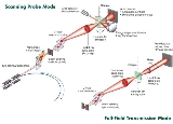
The hard X-ray nanoprobe at the Center for Nanoscale Materials
(CNM), Argonne National Lab advanced the state of the art by providing a hard X-ray microscopy beamline with the highest spatial resolution in the world. It provides for fluorescence, diffraction, and transmission imaging with hard X-rays at a spatial resolution of 30 nm or better. A dedicated source, beamline, and optics form the basis for these capabilities. This unique instrument is not only key to the specific research areas of the CNM; it is also be of general utility to the broader nanoscience community in studying nanomaterials and nanostructures, particularly for embedded structures.
The combination of diffraction, fluorescence, and transmission contrast in a single tool provides unique characterization capabilities for nanoscience. Current hard X-ray microprobes based on Fresnel zone plate optics have demonstrated a spatial resolution of 150 nm at a photon energy of 8-10 keV. With advances in the fabrication of zone plate optics, coupled with an optimized beamline design, the performance goal is a spatial resolution of 30 nm. The nanoprobe covers the spectral range of 3-30 keV, and the working distance between the focusing optics and the sample are typically in the range of 10–20 mm.

. In this mode, either attenuation or phase shift of the X-ray beam by the sample can be measured. Absorption contrast can be used to map the sample’s density. Particular elemental constituents can be located using measurements on each side of an absorption edge to give an element-specific difference image with moderate sensitivity. Phase-contrast imaging can be sensitive to internal structure even when absorption is low and can be enhanced by tuning the X-ray energy.
Diffraction
. By measuring X-rays diffracted from the sample, one can obtain local structural information, such as crystallographic phase, strain, and texture, with an accuracy 100 times higher than with standard electron diffraction
.
Fluorescence
. Induced X-ray fluorescence reveals the spatial distribution of individual elements in a sample. Because an X-ray probe offers 1,000 times higher sensitivity than electron probes, the fluorescence technique is a powerful tool for quantitative trace element analysis, important for understanding material properties such as second-phase particles, defects, and interfacial segregation.
Spectroscopy
. In spectroscopy mode, the primary X-ray beam’s energy is scanned across the absorption edge of an element, providing information on its chemical state (XANES
) or its local environment (EXAFS), which allows the study of disordered samples.
Polarization. Both linearly and circularly polarized X-rays will be available. Contrast due to polarization is invaluable in distinguishing fluorescence and diffraction signals and imaging magnetic domain structure by using techniques such as linear and circular dichroism and magnetic diffraction.
Tomography
. In X-ray tomography, one of these modes is combined with sample rotation to produce a series of two-dimensional projection images, to be used for reconstructing the sample’s internal three-dimensional structure. This will be particularly important for observing the morphology of complex nanostructures.
In summary, a hard X-ray nanoprobe provides advantages such as being noninvasive and quantitative, requiring minimal sample preparation, giving sub-optical spatial resolution, having the ability to penetrate inside a sample and study its internal structure, and having enhanced ability to study processes in situ. Another important distinction from charged-particle probes is that X-rays do not interact with applied electric or magnetic fields, which is an advantage for in-field studies. The design of the nanoprobe beamline aims to preserve these potential advantages.
Center for Nanoscale Materials
The Center for Nanoscale Materials is one of five Nanoscale Science Research Centers the United States Department of Energy sponsors. The Center is at Argonne National Laboratory location in Argonne, Illinois....
(CNM), Argonne National Lab advanced the state of the art by providing a hard X-ray microscopy beamline with the highest spatial resolution in the world. It provides for fluorescence, diffraction, and transmission imaging with hard X-rays at a spatial resolution of 30 nm or better. A dedicated source, beamline, and optics form the basis for these capabilities. This unique instrument is not only key to the specific research areas of the CNM; it is also be of general utility to the broader nanoscience community in studying nanomaterials and nanostructures, particularly for embedded structures.
The combination of diffraction, fluorescence, and transmission contrast in a single tool provides unique characterization capabilities for nanoscience. Current hard X-ray microprobes based on Fresnel zone plate optics have demonstrated a spatial resolution of 150 nm at a photon energy of 8-10 keV. With advances in the fabrication of zone plate optics, coupled with an optimized beamline design, the performance goal is a spatial resolution of 30 nm. The nanoprobe covers the spectral range of 3-30 keV, and the working distance between the focusing optics and the sample are typically in the range of 10–20 mm.
Modes of operation
TransmissionTransmittance
In optics and spectroscopy, transmittance is the fraction of incident light at a specified wavelength that passes through a sample. A related term is absorptance, or absorption factor, which is the fraction of radiation absorbed by a sample at a specified wavelength...
. In this mode, either attenuation or phase shift of the X-ray beam by the sample can be measured. Absorption contrast can be used to map the sample’s density. Particular elemental constituents can be located using measurements on each side of an absorption edge to give an element-specific difference image with moderate sensitivity. Phase-contrast imaging can be sensitive to internal structure even when absorption is low and can be enhanced by tuning the X-ray energy.
Diffraction
Diffraction
Diffraction refers to various phenomena which occur when a wave encounters an obstacle. Italian scientist Francesco Maria Grimaldi coined the word "diffraction" and was the first to record accurate observations of the phenomenon in 1665...
. By measuring X-rays diffracted from the sample, one can obtain local structural information, such as crystallographic phase, strain, and texture, with an accuracy 100 times higher than with standard electron diffraction
Electron diffraction
Electron diffraction refers to the wave nature of electrons. However, from a technical or practical point of view, it may be regarded as a technique used to study matter by firing electrons at a sample and observing the resulting interference pattern...
.
Fluorescence
Fluorescence
Fluorescence is the emission of light by a substance that has absorbed light or other electromagnetic radiation of a different wavelength. It is a form of luminescence. In most cases, emitted light has a longer wavelength, and therefore lower energy, than the absorbed radiation...
. Induced X-ray fluorescence reveals the spatial distribution of individual elements in a sample. Because an X-ray probe offers 1,000 times higher sensitivity than electron probes, the fluorescence technique is a powerful tool for quantitative trace element analysis, important for understanding material properties such as second-phase particles, defects, and interfacial segregation.
Spectroscopy
Spectroscopy
Spectroscopy is the study of the interaction between matter and radiated energy. Historically, spectroscopy originated through the study of visible light dispersed according to its wavelength, e.g., by a prism. Later the concept was expanded greatly to comprise any interaction with radiative...
. In spectroscopy mode, the primary X-ray beam’s energy is scanned across the absorption edge of an element, providing information on its chemical state (XANES
XANES
X-ray Absorption Near Edge Structure , also known as Near edge X-ray absorption fine structure is a type of absorption spectroscopy. NEXAFS also at times used the abbreviation EXAFS....
) or its local environment (EXAFS), which allows the study of disordered samples.
Polarization. Both linearly and circularly polarized X-rays will be available. Contrast due to polarization is invaluable in distinguishing fluorescence and diffraction signals and imaging magnetic domain structure by using techniques such as linear and circular dichroism and magnetic diffraction.
Tomography
Tomography
Tomography refers to imaging by sections or sectioning, through the use of any kind of penetrating wave. A device used in tomography is called a tomograph, while the image produced is a tomogram. The method is used in radiology, archaeology, biology, geophysics, oceanography, materials science,...
. In X-ray tomography, one of these modes is combined with sample rotation to produce a series of two-dimensional projection images, to be used for reconstructing the sample’s internal three-dimensional structure. This will be particularly important for observing the morphology of complex nanostructures.
In summary, a hard X-ray nanoprobe provides advantages such as being noninvasive and quantitative, requiring minimal sample preparation, giving sub-optical spatial resolution, having the ability to penetrate inside a sample and study its internal structure, and having enhanced ability to study processes in situ. Another important distinction from charged-particle probes is that X-rays do not interact with applied electric or magnetic fields, which is an advantage for in-field studies. The design of the nanoprobe beamline aims to preserve these potential advantages.
Activities
- Hard X-ray nanoprobe
- Large numerical aperture optics for hard X-rays
- Time-resolved, stroboscopic measurements
- Full-field imaging
- In situ studies of nanomaterials growth processes
- Scanning probe fluorescenceFluorescenceFluorescence is the emission of light by a substance that has absorbed light or other electromagnetic radiation of a different wavelength. It is a form of luminescence. In most cases, emitted light has a longer wavelength, and therefore lower energy, than the absorbed radiation...
, diffractionDiffractionDiffraction refers to various phenomena which occur when a wave encounters an obstacle. Italian scientist Francesco Maria Grimaldi coined the word "diffraction" and was the first to record accurate observations of the phenomenon in 1665...
, and transmissionTransmittanceIn optics and spectroscopy, transmittance is the fraction of incident light at a specified wavelength that passes through a sample. A related term is absorptance, or absorption factor, which is the fraction of radiation absorbed by a sample at a specified wavelength...
phase contrast imagingPhase contrast microscopyPhase contrast microscopy is an optical microscopy illumination technique of great importance to biologists in which small phase shifts in the light passing through a transparent specimen are converted into amplitude or contrast changes in the image.A phase contrast microscope does not require... - Polarization dependent scattering
- General nanomaterials characterization with X-rays, including small-angle scatteringSmall-angle scatteringSmall-angle scattering is a scattering technique based on the deflection of a beam of particles, or an electromagnetic or acoustic wave, away from the straight trajectory after it interacts with structures that are much larger than the wavelength of the radiation. The deflection is small hence...
(SAXS)