

Vision restoration therapy
Encyclopedia
Vision Restoration Therapy (VRT) is a noninvasive, nonsurgical form of Vision therapy
provided by NovaVision. This therapy was developed by Bernhard Sabel, Ph.D. The phenomenon underlying the therapy is visual neuroplasticity
.
defects regain function by stimulating existing visual neurons to rewire and make new connections. This therapy is cleared by the U.S. Food and Drug Administration
(FDA) to treat visual field defects due to stroke
, head injury, brain tumors, and brain surgery. Some other defects that can also be treated with VRT include hemianopia, quadrantanopia
, scotoma
, and diffuse field defect.
occurs concurrently with reorganization of the visual system
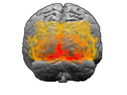
by new connections made by stimulated neurons. Using the imaging technique of fMRI, it was found that brain activity was altered after Vision Restoration Therapy. This associates cerebral reorganization with VRT treatment.
The cerebrum is involved with higher brain function, and one component of the cerebrum is the primary visual cortex. The primary visual cortex is a region in the occipital lobe
that can be altered by neuroplasticity to create new neuronal pathways around damaged areas to help regain lost visual functions. Sensory visual information is sent from the retina of the eye to the Lateral geniculate nucleus
(LGN) in the Thalamus
, which relays the visual information to the primary visual cortex by the fibers of the optic radiation
. Lesions or damage to parts of the brain that cause visual field defects usually occur posterior to the optic chiasm
. Although the exact mechanisms that underlie regaining visual field functions through visual neuroplasticity and VRT are not yet fully known, the reorganization of the primary visual cortex is thought to make new connections and pathways in the optic radiation to the LGN to help regain visual field functions. The stimulation of existing neurons near a damaged site in the brain can form new synapse
s with other functional neurons to help take on and compensate for the function lost due to the damaged neurons. This is what is theorized to occur during VRT treatment.
Vision Restoration Therapy stimulates the retina of the eye using repetitive points of light that flash on a computer screen. These flashing lights are aimed to stimulate the border of the blind area of a patient’s visual field. The repetitive stimulation is used to help promote visual neuroplasticity and ultimately make new neuronal connections to regain and expand the visual field.
Patients perform this therapy using the VRT device as stated before. The VRT device consists of a chin rest and a specialized computer screen that has High Resolution Perimetry (HRP) incorporated into its software. The built-in HRP helps in assessing and tracking the progress of the patient. The device sends this data directly to the therapy provider for feedback to modify the treatment if necessary. HRP is used to map the visual field of a patient. This allows the therapy providers to have a representation of the patient's visual field. For a general VRT session, the patient’s head is situated at eye level with the VRT device. The therapy begins by projecting a fixation point in the center of the screen. The patient focuses on this fixation point for the entire session. As the patient is focusing on the central fixation point, an individual point appears somewhere on the screen. Only one point is projected at a time alongside the fixation point. The patient must click the mouse every time he or she sees this point while focusing on the fixation point. These responses are collected to calculate stimuli detection rate and response time of the patient. In an effort to see the points on the screen most clearly, the therapy should be conducted in a dimly lit room.
When VRT was first introduced, the primary skepticism involved the data collection methods that the VRT researchers used to quantify the effective results of using the therapy. The main argument was that the HRP data from the VRT device that was used to measure the progress of the patients was the same data used to show the effectiveness of VRT. These data would not be as reliable as data gathered using some other Perimetry
technique. Skeptics turned to studies that assessed VRT effectiveness using Tubinger Automatic Perimetry and Scanning laser ophthalmoscopy
(SLO), which showed that no beneficial results in improving the visual field were associated with VRT use. Skepticism also arose about the quality of life questionnaire surveys that patients took after VRT treatment. The possibility of the placebo effect
could be present in the answering of these questionnaires. Patients could have stated that their qualities of life did improve just because they had finished the therapy, even if no improvements actually occurred. Others questioned the neuroplastic mechanism behind VRT, stating that no salvageable tissue remains in the occipital lobe with vision deficits such as hemianopia. Neuroplasticity cannot make new connections according to this claim, which debunks VRT in its entirety. It was proclaimed that eye movements were the cause of visual rehabilitation instead of self regeneration of the brain and visual neuroplasticity.
techniques in Vision Restoration Therapy effectiveness. To reiterate, perimetry is visual field testing, and there are several perimetry techniques to measure visual fields. It was found that perimetric analysis using SLO was a more difficult task than commonly used Perimetry techniques such as HRP. For comparison, SLO and HRP will be used as examples. SLO displays 3 black points on a bright red background, whereas HRP uses a bright red point on a black or grey background. The task of perceiving the black points on the red background in SLO was found to be a difficult task for patients with brain damage. Also, the added attention needed to perceive all three points in SLO makes the task more difficult. Lastly, patients acknowledge that they see the points of light verbally in SLO. Patients just click the mouse after seeing a point in HRP. Verbal acknowledgement may reduce response time and seems an inefficient way to measure response time. These findings challenged the skeptics in relying on SLO data to measure the effectiveness of VRT. A recent study assessed VRT effectiveness using fundus-controlled microperimetry to measure visual fields. The findings concluded that VRT was indeed effective in expanding the visual field. The microperimetry technique compensates for eye movements.
is beneficial for practitioners to get a general idea of the potential improvements by a certain procedure or treatment. Researchers have developed a Treatment Outcome Predictive Model to calculate the potential visual field recovery after VRT. This model uses several perimetric variables to make scientific predictions on a patient’s potential visual field recovery using VRT.
and glaucoma
. Although these are pilot studies and require more data, it seems promising that VRT can help restore some visual functions of patients with anterior ischemic optic neuropathy. The same promising results were found for patients with glaucoma. Some visual functions were able to be restored after VRT treatment.
Vision therapy
Vision therapy, also known as visual training, vision training, or visual therapy, is a broad group of techniques aimed at correcting and improving binocular, oculomotor, visual processing, and perceptual disorders."-Historical development:...
provided by NovaVision. This therapy was developed by Bernhard Sabel, Ph.D. The phenomenon underlying the therapy is visual neuroplasticity
Neuroplasticity
Neuroplasticity is a non-specific neuroscience term referring to the ability of the brain and nervous system in all species to change structurally and functionally as a result of input from the environment. Plasticity occurs on a variety of levels, ranging from cellular changes involved in...
.
Description of therapy
Vision Restoration Therapy is a computer-based treatment therapy, and is used to help patients with visual fieldVisual field
The term visual field is sometimes used as a synonym to field of view, though they do not designate the same thing. The visual field is the "spatial array of visual sensations available to observation in introspectionist psychological experiments", while 'field of view' "refers to the physical...
defects regain function by stimulating existing visual neurons to rewire and make new connections. This therapy is cleared by the U.S. Food and Drug Administration
Food and Drug Administration
The Food and Drug Administration is an agency of the United States Department of Health and Human Services, one of the United States federal executive departments...
(FDA) to treat visual field defects due to stroke
Stroke
A stroke, previously known medically as a cerebrovascular accident , is the rapidly developing loss of brain function due to disturbance in the blood supply to the brain. This can be due to ischemia caused by blockage , or a hemorrhage...
, head injury, brain tumors, and brain surgery. Some other defects that can also be treated with VRT include hemianopia, quadrantanopia
Quadrantanopia
Quadrantanopia, quadrantanopsia, or quadrant anopia refers to an anopia affecting a quarter of the field of vision.It can be associated with a lesion of an optic radiation...
, scotoma
Scotoma
A scotoma is an area of partial alteration in one's field of vision consisting of a partially diminished or entirely degenerated visual acuity which is surrounded by a field of normal - or relatively well-preserved - vision.Every normal mammalian eye has a scotoma in its field of vision, usually...
, and diffuse field defect.
Criteria to undergo therapy
To have the greatest chance of possible recovery, patients must first go through screening criteria with their physicians to see if they can effectively undergo VRT. A few of the major criteria are listed below.- Patients that have photosensitive epilepsyPhotosensitive epilepsyPhotosensitive epilepsy is a form of epilepsy in which seizures are triggered by visual stimuli that form patterns in time or space, such as flashing lights, bold, regular patterns, or regular moving patterns.-Symptoms:...
, serious eye disease, or prominent cognitive deficitCognitive deficitCognitive deficit, also known as cognitive impairment is an inclusive term to describe any characteristic that acts as a barrier to cognitive performance...
s may not be eligible to undergo VRT treatment. - An appropriate level of cognition is necessary due to the demand of great concentration and attention needed to pursue an effective treatment regimen.
- A minimum visual acuityVisual acuityVisual acuity is acuteness or clearness of vision, which is dependent on the sharpness of the retinal focus within the eye and the sensitivity of the interpretative faculty of the brain....
of 20/200 is also required for proper stimuliStimulus (physiology)In physiology, a stimulus is a detectable change in the internal or external environment. The ability of an organism or organ to respond to external stimuli is called sensitivity....
detection.
Advantages
Listed below are some of the advantages associated with VRT.- The benefits of the therapy for a patient are not confined by the duration of time that the patient has had the visual defect. In other words, the benefits are not limited by the time difference between the time the patient developed the defect and the time of starting Vision Restoration Therapy.
- There is no age limitation for a patient to undergo treatment.
- The type of visual field defect does not limit the benefits of VRT.
- There are no major side effects associated with VRT treatment. A few patients have reported mild infrequent headaches during treatment.
Statistics
It is reported that about 70% of the patients that undergo the treatment have improvements in their visual fields. About 90% of the patients also stated that the use of the therapy has been accompanied by at least one major benefit. Some of these benefits include, improved reading, improved mobility, increased confidence, and independence. Around 88% of the patients also reported a better quality of life after completion of the therapy. Although results vary among patients, the therapy expands the visual field on average by 5°. This is associated with a 5° retraction in the blind field. The blind field is the antagonist of the visual field, and an expansion of the blind field is associated with visual field loss.Neuroplasticity
The discovery of neuroplasticity has been hailed by some scientists to be one of the greatest discoveries ever. Neuroplasticity is the ability of the brain to adapt to the environment by making changes in itself over time. New technologies and therapies are being developed to harness the power of neuroplasticity in order to heal the repercussions of brain-related damage and injury. Vision Restoration Therapy is one such therapy that exploits visual neuroplasticity to alleviate visual field defects. VRT has been shown to expand the visual fields of patients and the repetitive activation and stimulation of functional neurons is the heart of the therapy.Harnessing the power of visual neuroplasticity for therapeutic purposes
Work in understanding visual neuroplasticity has made progress in the past decade. The potential for visual neuroplasticity can exist for a long duration of time. There are two approaches as of now to take advantage of this potential visual neuroplasticity.- The first approach involves rescuing, regenerating, or transplanting visual neurons by biological/pharmacological methods. One example of this approach is the discovery of the regeneration of axons of retinal ganglion cells (RGCs) by introducing several proteins into the chemical environment of these cells. Some of these known proteins that induce axon growth of RGCs include lamininLamininLaminins are major proteins in the basal lamina , a protein network foundation for most cells and organs...
, Gap-43 proteinGap-43 proteinGrowth Associated Protein 43 also known as Gap43 is a protein that in humans is encoded by the GAP43 gene.Gap43 has been termed a 'growth' or 'plasticity' protein because it is expressed at high levels in neuronal growth cones during development, during axonal regeneration and is phosphorylated...
, fatty acid binding protein, calmodulinCalmodulinCalmodulin is a calcium-binding protein expressed in all eukaryotic cells...
, Alpha crystallinAlpha crystallinAlpha crystallin is a water-soluble structural protein found in the lens of the eye. Compared to the other crystallins, it elutes earlier from a gel filtration chromatography column....
, IFN-gamma, cyclin-dependent kinase inhibitor proteinCyclin-dependent kinase inhibitor proteinA cyclin-dependent kinase inhibitor protein is a protein which inhibits cyclin-dependent kinase. Several function as tumor suppressor genes. Cell cycle progression is negatively controlled by cyclin-dependent kinases inhibitors . CDIs are involved in cell cycle arrest at the G1 phase.-Examples:...
, beta-hemoglobin, 60s-ribosomal protein, GAP-DH, and ADP-ribosylation factor. - The second approach involves improving visual functions by stimulating the associated neurons in either the blind region of the visual field through different pathways or by stimulating damaged areas of the border region. This approach is a more promising approach than the first in harnessing visual neuroplasticity to help regain visual functions. Vision Restoration Therapy is an example of this approach.
Biology of visual neuroplasticity
With visual neuroplasticity, reorganization of the physical structure of the brainBrain
The brain is the center of the nervous system in all vertebrate and most invertebrate animals—only a few primitive invertebrates such as sponges, jellyfish, sea squirts and starfishes do not have one. It is located in the head, usually close to primary sensory apparatus such as vision, hearing,...
occurs concurrently with reorganization of the visual system
Visual system
The visual system is the part of the central nervous system which enables organisms to process visual detail, as well as enabling several non-image forming photoresponse functions. It interprets information from visible light to build a representation of the surrounding world...
by new connections made by stimulated neurons. Using the imaging technique of fMRI, it was found that brain activity was altered after Vision Restoration Therapy. This associates cerebral reorganization with VRT treatment.
The cerebrum is involved with higher brain function, and one component of the cerebrum is the primary visual cortex. The primary visual cortex is a region in the occipital lobe
Occipital lobe
The occipital lobe is the visual processing center of the mammalian brain containing most of the anatomical region of the visual cortex. The primary visual cortex is Brodmann area 17, commonly called V1...
that can be altered by neuroplasticity to create new neuronal pathways around damaged areas to help regain lost visual functions. Sensory visual information is sent from the retina of the eye to the Lateral geniculate nucleus
Lateral geniculate nucleus
The lateral geniculate nucleus is the primary relay center for visual information received from the retina of the eye. The LGN is found inside the thalamus of the brain....
(LGN) in the Thalamus
Thalamus
The thalamus is a midline paired symmetrical structure within the brains of vertebrates, including humans. It is situated between the cerebral cortex and midbrain, both in terms of location and neurological connections...
, which relays the visual information to the primary visual cortex by the fibers of the optic radiation
Optic radiation
The optic radiation is a collection of axons from relay neurons in the lateral geniculate nucleus of the thalamus carrying visual information to the visual cortex along the calcarine fissure.There is one such tract on each side of the brain.-Parts:A distinctive...
. Lesions or damage to parts of the brain that cause visual field defects usually occur posterior to the optic chiasm
Optic chiasm
The optic chiasm or optic chiasma is the part of the brain where the optic nerves partially cross...
. Although the exact mechanisms that underlie regaining visual field functions through visual neuroplasticity and VRT are not yet fully known, the reorganization of the primary visual cortex is thought to make new connections and pathways in the optic radiation to the LGN to help regain visual field functions. The stimulation of existing neurons near a damaged site in the brain can form new synapse
Synapse
In the nervous system, a synapse is a structure that permits a neuron to pass an electrical or chemical signal to another cell...
s with other functional neurons to help take on and compensate for the function lost due to the damaged neurons. This is what is theorized to occur during VRT treatment.
Vision Restoration Therapy stimulates the retina of the eye using repetitive points of light that flash on a computer screen. These flashing lights are aimed to stimulate the border of the blind area of a patient’s visual field. The repetitive stimulation is used to help promote visual neuroplasticity and ultimately make new neuronal connections to regain and expand the visual field.
Process of therapy
Patients undergo therapy in the comforts of their home due to it being a software-based therapy sent to patients as a specialized portable apparatus called NovaVision VRT Device. The therapy requires a prescription from a physician in order to begin. A set of initial evaluation tests are taken using the VRT device to assess the amount of visual impairment and the locations of the borders of the visual field deficit. The therapy provider, NovaVision, then analyzes these data to customize the treatment to target these borders of deficit to help expand the visual field. Usually, patients complete the treatment course in 6 months. The therapy consists of patients using the VRT device for 30 minutes twice a day for 6 days of the week during the 6 month treatment course.Patients perform this therapy using the VRT device as stated before. The VRT device consists of a chin rest and a specialized computer screen that has High Resolution Perimetry (HRP) incorporated into its software. The built-in HRP helps in assessing and tracking the progress of the patient. The device sends this data directly to the therapy provider for feedback to modify the treatment if necessary. HRP is used to map the visual field of a patient. This allows the therapy providers to have a representation of the patient's visual field. For a general VRT session, the patient’s head is situated at eye level with the VRT device. The therapy begins by projecting a fixation point in the center of the screen. The patient focuses on this fixation point for the entire session. As the patient is focusing on the central fixation point, an individual point appears somewhere on the screen. Only one point is projected at a time alongside the fixation point. The patient must click the mouse every time he or she sees this point while focusing on the fixation point. These responses are collected to calculate stimuli detection rate and response time of the patient. In an effort to see the points on the screen most clearly, the therapy should be conducted in a dimly lit room.
Skepticism
The effectiveness of VRT has been a controversial topic. Beneficial results have been researched and documented by testing small groups of patients. This has led to skepticism by some of the scientific community. Even though VRT has been available for a few years now, optometrists, ophthalmologists, and other eye specialists do not regularly recommend VRT for their patients. The primary cause of this is that eye specialists rarely recommend this therapy to other eye specialists. The skepticism of VRT may be the source of the lack of recommendation.When VRT was first introduced, the primary skepticism involved the data collection methods that the VRT researchers used to quantify the effective results of using the therapy. The main argument was that the HRP data from the VRT device that was used to measure the progress of the patients was the same data used to show the effectiveness of VRT. These data would not be as reliable as data gathered using some other Perimetry
Perimetry
Perimetry or campimetry is the systematic measurement of differential light sensitivity in the visual field by the detection of the presence of test targets on a defined background. Visual field testing can be performed clinically with confrontational field testing keeping the subject's gaze fixed...
technique. Skeptics turned to studies that assessed VRT effectiveness using Tubinger Automatic Perimetry and Scanning laser ophthalmoscopy
Scanning laser ophthalmoscopy
Scanning laser ophthalmoscopy is a method of examination of the eye. It uses the technique of confocal laser scanning microscopy for diagnostic imaging of retina or cornea of the human eye....
(SLO), which showed that no beneficial results in improving the visual field were associated with VRT use. Skepticism also arose about the quality of life questionnaire surveys that patients took after VRT treatment. The possibility of the placebo effect
Placebo effect
Placebo effect may refer to:* Placebo effect, the tendency of any medication or treatment, even an inert or ineffective one, to exhibit results simply because the recipient believes that it will work...
could be present in the answering of these questionnaires. Patients could have stated that their qualities of life did improve just because they had finished the therapy, even if no improvements actually occurred. Others questioned the neuroplastic mechanism behind VRT, stating that no salvageable tissue remains in the occipital lobe with vision deficits such as hemianopia. Neuroplasticity cannot make new connections according to this claim, which debunks VRT in its entirety. It was proclaimed that eye movements were the cause of visual rehabilitation instead of self regeneration of the brain and visual neuroplasticity.
Clinical studies and research
Much of the research that was initially conducted did not convince many of the scientific community that VRT is an effective treatment option for large scale use. As a result, many studies have been conducted to target the arguments of the skeptics.Studies with large sample sizes
Much skepticism arose due to the fact that many of the initial studies of VRT effectiveness used only small sample sizes. One study aimed to show the effectiveness of VRT using a large sample size as a result. Researchers assessed the effectiveness of VRT by testing 302 patients. The study used the qualitative data from HRP, the stimulus detection rate, and the average response time before and after the patients underwent VRT. The stimulus detection rate did vary. About 38.3% of patients had great improvements, 32.6% had moderate improvement, and 29.1% had no improvement. The average reaction time also improved for the patients. It fell from 463.6 ± 5.08 ms to 446.2 ± 5.23 ms. This study showed that even assessing a large sample size yields the same positive results of effectiveness as the small sample size studies.Comparing perimetry techniques
The other criticism was that the perimetric data from the SLO showed that there was no improvement in the visual fields of patients that undergo VRT. Studies have compared and contrasted the various PerimetryPerimetry
Perimetry or campimetry is the systematic measurement of differential light sensitivity in the visual field by the detection of the presence of test targets on a defined background. Visual field testing can be performed clinically with confrontational field testing keeping the subject's gaze fixed...
techniques in Vision Restoration Therapy effectiveness. To reiterate, perimetry is visual field testing, and there are several perimetry techniques to measure visual fields. It was found that perimetric analysis using SLO was a more difficult task than commonly used Perimetry techniques such as HRP. For comparison, SLO and HRP will be used as examples. SLO displays 3 black points on a bright red background, whereas HRP uses a bright red point on a black or grey background. The task of perceiving the black points on the red background in SLO was found to be a difficult task for patients with brain damage. Also, the added attention needed to perceive all three points in SLO makes the task more difficult. Lastly, patients acknowledge that they see the points of light verbally in SLO. Patients just click the mouse after seeing a point in HRP. Verbal acknowledgement may reduce response time and seems an inefficient way to measure response time. These findings challenged the skeptics in relying on SLO data to measure the effectiveness of VRT. A recent study assessed VRT effectiveness using fundus-controlled microperimetry to measure visual fields. The findings concluded that VRT was indeed effective in expanding the visual field. The microperimetry technique compensates for eye movements.
Role of eye movements on visual rehabilitation
Studies were initiated to observe the role of eye movements as the reason behind visual field improvements. It has been debated that patients with visual field defects try to make up for their limited vision by making more frequent eye movements in an effort to expand their visual field. This theory debunks the efficacy of VRT and neuroplasticity as the cause of visual field expansion. Research has found that VRT does not change eye movements and the visual field improvements and expansion are independent of these movements.Long-term use and stability of VRT
Other studies have looked into long term use of VRT and the stability of visual field improvements after VRT treatment. One study looked into the effectiveness of VRT in improving visual fields by using the therapy for 12 months as opposed to the recommended 6 months. It was found that miniscule improvements occur after the first 6 month interval. The result also validate that treatment for 6 months is the optimal time for the therapy. Another study looked into the stability of the results from VRT treatment. Patients were reassessed using Perimetry techniques about 2 years after they underwent VRT. The findings suggest that there is stability in the visual field improvements after VRT treatment. However, the stability is dependent on the patient to use the restored visual functions continuously and naturally to keep stimulating the new neuronal connections to achieve stronger visual functioning.Predictive outcome models of VRT effectiveness
Having a predictive modelPredictive modelling
Predictive modelling is the process by which a model is created or chosen to try to best predict the probability of an outcome. In many cases the model is chosen on the basis of detection theory to try to guess the probability of an outcome given a set amount of input data, for example given an...
is beneficial for practitioners to get a general idea of the potential improvements by a certain procedure or treatment. Researchers have developed a Treatment Outcome Predictive Model to calculate the potential visual field recovery after VRT. This model uses several perimetric variables to make scientific predictions on a patient’s potential visual field recovery using VRT.
Pilot studies
Pilot studies have also looked into the effects of Vision Restoration Therapy in treating visual field defects that have resulted from anterior ischemic optic neuropathyAnterior ischemic optic neuropathy
Anterior ischemic optic neuropathy is a medical condition involving loss of vision due to damage to the optic nerve from insufficient blood supply. AION is generally divided into two types: arteritic AION and non-arteritic AION...
and glaucoma
Glaucoma
Glaucoma is an eye disorder in which the optic nerve suffers damage, permanently damaging vision in the affected eye and progressing to complete blindness if untreated. It is often, but not always, associated with increased pressure of the fluid in the eye...
. Although these are pilot studies and require more data, it seems promising that VRT can help restore some visual functions of patients with anterior ischemic optic neuropathy. The same promising results were found for patients with glaucoma. Some visual functions were able to be restored after VRT treatment.