Ventricular escape beat
Encyclopedia

Ventricle (heart)
In the heart, a ventricle is one of two large chambers that collect and expel blood received from an atrium towards the peripheral beds within the body and lungs. The Atria primes the Pump...
of the heart
Heart
The heart is a myogenic muscular organ found in all animals with a circulatory system , that is responsible for pumping blood throughout the blood vessels by repeated, rhythmic contractions...
; normally the heart rhythm is begun in the atria
Atria
Atria may refer to:*Atrium , an anatomical structure of the heart*Atrium , a large open space within a building*Atria or Alpha Trianguli Australis, a star in the constellation Triangulum Australe...
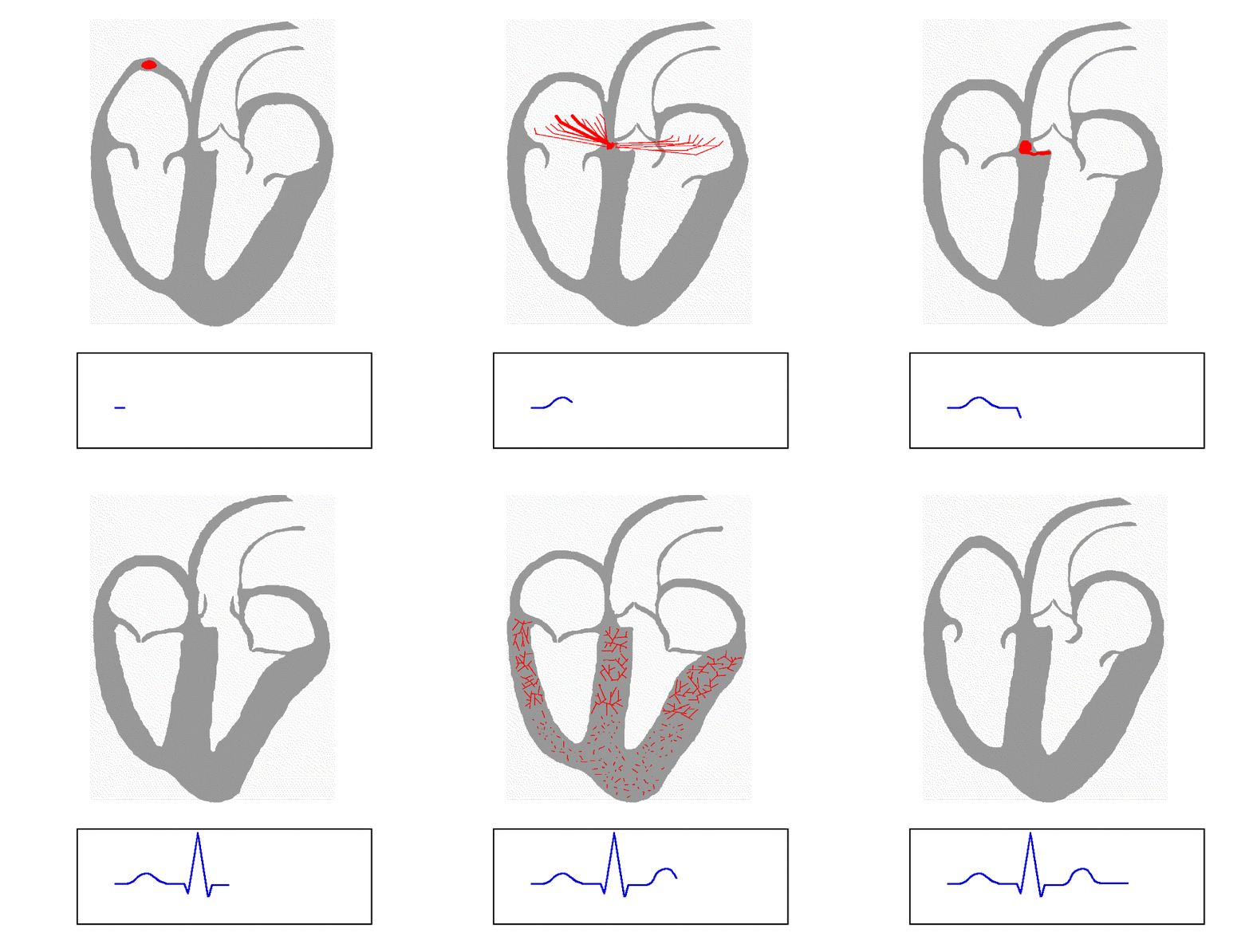
of the heart and is subsequently transmitted to the ventricles. The ventricular escape beat follows a long pause in ventricular rhythm and acts to prevent cardiac arrest
Cardiac arrest
Cardiac arrest, is the cessation of normal circulation of the blood due to failure of the heart to contract effectively...
. It indicates a failure of the electrical conduction system of the heart
Electrical conduction system of the heart
The normal intrinsic electrical conduction of the heart allows electrical propagation to be transmitted from the Sinoatrial Node through both atria and forward to the Atrioventricular Node. Normal/baseline physiology allows further propagation from the AV node to the ventricle or Purkinje Fibers...
to stimulate the ventricles (which would lead to the absence of heartbeats, unless ventricular escape beats occur).
Causes
Ventricular escape beats occur when the rate of electrical discharge reaching the ventricles (normally initiated by the heart's sinoatrial nodeSinoatrial node
The sinoatrial node is the impulse-generating tissue located in the right atrium of the heart, and thus the generator of normal sinus rhythm. It is a group of cells positioned on the wall of the right atrium, near the entrance of the superior vena cava...
(SA node), transmitted to the atrioventricular node
Atrioventricular node
The atrioventricular node is a part of the electrical control system of the heart that coordinates heart rate. It electrically connects atrial and ventricular chambers...
(AV node), and then further transmitted to the ventricles) falls below the base rate determined by the ventricular pacemaker cells.
An escape beat usually occurs 2–3 seconds after an electrical impulse has failed to reach the ventricles
This phenomenon can be caused by the sinoatrial node
Sinoatrial node
The sinoatrial node is the impulse-generating tissue located in the right atrium of the heart, and thus the generator of normal sinus rhythm. It is a group of cells positioned on the wall of the right atrium, near the entrance of the superior vena cava...
(SA node) failing to initiate a beat, by a failure of the conductivity from the SA node to the atrioventricular node
Atrioventricular node
The atrioventricular node is a part of the electrical control system of the heart that coordinates heart rate. It electrically connects atrial and ventricular chambers...
(AV node), or by atrioventricular block
Atrioventricular block
An atrioventricular block involves the impairment of the conduction between the atria and ventricles of the heart.The causes of pathological AV block are varied and include ischaemia, infarction, fibrosis or drugs. Certain AV blocks can also be found as normal variants, such as in athletes or...
(especially third degree AV block). Normally, the pacemaker cells of the sinoatrial node discharge at the highest frequency and are thus dominant over other cells with pacemaker activity. The AV node normally has the second fastest discharge rate. When the sinus rate falls below the discharge rate of the AV node, this becomes the dominant pacemaker, and the result is called a junctional escape beat
Junctional escape beat
A junctional escape beat is a delayed heartbeat originating not from the atrium but from an ectopic focus somewhere in the AV junction. It occurs when the rate of depolarization of the sinoatrial node falls below the rate of the atrioventricular node. This dysrhythmia also may occur when the...
. If the rate from both the SA and AV node fall below the discharge rate of ventricular pacemaker cells, a ventricular escape beat ensues.
An escape beat is a form of cardiac arrhythmia, in this case known as an ectopic beat
Cardiac ectopy
Ectopic beat is a disturbance of the cardiac rhythm frequently related to the electrical conduction system of the heart, in which beats arise from fiber or group of fibers outside the region in the heart muscle ordinarily responsible for impulse formation, i.e., the Sinus node...
. It can be considered a form of ectopic pacemaker activity that is unveiled by lack of other pacemakers to stimulate the ventricles. Ventricular pacemaker cells discharge at a slower rate than the SA or AV node. While the SA node typically initiates a rate of 70 beats per minute (BPM), the atrioventricular node
Atrioventricular node
The atrioventricular node is a part of the electrical control system of the heart that coordinates heart rate. It electrically connects atrial and ventricular chambers...
(AV node) is usually only capable of generating a rhythm at 40-60 BPM or less. Ventricular contraction rate is thus reduced by 15-40 beats per minute.
If there are only one or two ectopic beats, they are considered escape beats. If this causes a semi-normal rhythm to arise it is considered an idioventricular rhythm
Idioventricular rhythm
Normally, the pacemaker of the heart that is responsible for triggering each heart beat is the SA node. However, if the ventricle does not receive triggering signals at a rate high enough, the ventricular myocardium itself becomes the pacemaker . This is called Idioventricular Rhythm. The rate is...
.
The escape arrhythmia is a compensatory mechanism that indicates a serious underlying problem with the SA node or conduction system (commonly due to heart attack
Myocardial infarction
Myocardial infarction or acute myocardial infarction , commonly known as a heart attack, results from the interruption of blood supply to a part of the heart, causing heart cells to die...
or medication side effect
Adverse effect (medicine)
In medicine, an adverse effect is a harmful and undesired effect resulting from a medication or other intervention such as surgery.An adverse effect may be termed a "side effect", when judged to be secondary to a main or therapeutic effect. If it results from an unsuitable or incorrect dosage or...
), and because of its low rate, it can cause a drop in blood pressure
Blood pressure
Blood pressure is the pressure exerted by circulating blood upon the walls of blood vessels, and is one of the principal vital signs. When used without further specification, "blood pressure" usually refers to the arterial pressure of the systemic circulation. During each heartbeat, BP varies...
and syncope
Syncope (medicine)
Syncope , the medical term for fainting, is precisely defined as a transient loss of consciousness and postural tone characterized by rapid onset, short duration, and spontaneous recovery due to global cerebral hypoperfusion that most often results from hypotension.Many forms of syncope are...
.
Diagnosis
An electrocardiogramElectrocardiogram
Electrocardiography is a transthoracic interpretation of the electrical activity of the heart over a period of time, as detected by electrodes attached to the outer surface of the skin and recorded by a device external to the body...
can be used to identify a ventricular escape beat. The QRS portion of the electrocardiogram
Electrocardiogram
Electrocardiography is a transthoracic interpretation of the electrical activity of the heart over a period of time, as detected by electrodes attached to the outer surface of the skin and recorded by a device external to the body...
represents the ventricular depolarisation; in normal circumstances the QRS complex forms a sharp sudden peak. For a patient with a ventricular escape beat, the shape of the QRS complex is broader as the impulse can not travel quickly via the normal electrical conduction system.

Premature ventricular contraction
A premature ventricular contraction , also known as a premature ventricular complex, ventricular premature contraction , ventricular premature beat , or extrasystole, is a relatively common event where the heartbeat is initiated by the heart ventricles rather than by the sinoatrial node, the...
s (or premature ventricular contractions), which are spontaneous electrical discharges of the ventricles. These are not preceded by a pause; on the contrary they are often followed by a compensatory pause.
Cilostazol
Third degree AV block can be treated with Cilostazol which acts to increase Ventricular escape rateOuabain
OuabainOuabain
Ouabain which is also named g-strophanthin, is a poisonous cardiac glycoside.-Sources:Ouabain is found in the ripe seeds of African plants Strophanthus gratus and the bark of Acokanthera ouabaio.-Function:...
infusion decreases ventricular escape time and increases ventricular escape rhythm. However, a high dose of ouabain can lead to ventricular tachycardia
Tachycardia
Tachycardia comes from the Greek words tachys and kardia . Tachycardia typically refers to a heart rate that exceeds the normal range for a resting heart rate...
.

