

Two-dimensional gel electrophoresis
Encyclopedia
Gel electrophoresis
Gel electrophoresis is a method used in clinical chemistry to separate proteins by charge and or size and in biochemistry and molecular biology to separate a mixed population of DNA and RNA fragments by length, to estimate the size of DNA and RNA fragments or to separate proteins by charge...
commonly used to analyze proteins. Mixtures of proteins are separated by two properties in two dimensions on 2D gels.
Basis for separation
2-D electrophoresisElectrophoresis
Electrophoresis, also called cataphoresis, is the motion of dispersed particles relative to a fluid under the influence of a spatially uniform electric field. This electrokinetic phenomenon was observed for the first time in 1807 by Reuss , who noticed that the application of a constant electric...
begins with 1-D electrophoresis but then separates the molecules by a second property in a direction 90 degrees from the first. In 1-D electrophoresis, proteins (or other molecules) are separated in one dimension, so that all the proteins/molecules will lie along a lane but that the molecules are spread out across a 2-D gel. Because it is unlikely that two molecules will be similar in two distinct properties, molecules are more effectively separated in 2-D electrophoresis than in 1-D electrophoresis.
The two dimensions that proteins are separated into using this technique can be isoelectric point
Isoelectric point
The isoelectric point , sometimes abbreviated to IEP, is the pH at which a particular molecule or surface carries no net electrical charge....
, protein complex mass in the native state, and protein mass
Mass
Mass can be defined as a quantitive measure of the resistance an object has to change in its velocity.In physics, mass commonly refers to any of the following three properties of matter, which have been shown experimentally to be equivalent:...
.
To separate the proteins by isoelectric point is called isoelectric focusing
Isoelectric focusing
Isoelectric focusing , also known as electrofocusing, is a technique for separating different molecules by their electric charge differences...
(IEF). Thereby, a gradient of pH is applied to a gel and an electric potential is applied across the gel, making one end more positive than the other. At all pH values other than their isoelectric point, proteins will be charged. If they are positively charged, they will be pulled towards the more negative end of the gel and if they are negatively charged they will be pulled to the more positive end of the gel. The proteins applied in the first dimension will move along the gel and will accumulate at their isoelectric point; that is, the point at which the overall charge on the protein is 0 (a neutral charge).
For the analysis of the functioning of proteins in a cell
Cell (biology)
The cell is the basic structural and functional unit of all known living organisms. It is the smallest unit of life that is classified as a living thing, and is often called the building block of life. The Alberts text discusses how the "cellular building blocks" move to shape developing embryos....
, the knowledge of their cooperation is essential. Most often proteins act together in complexes to be fully functional. The analysis of this sub organelle
Organelle
In cell biology, an organelle is a specialized subunit within a cell that has a specific function, and is usually separately enclosed within its own lipid bilayer....
organisation of the cell requires techniques conserving the native state of the protein complex
Protein complex
A multiprotein complex is a group of two or more associated polypeptide chains. If the different polypeptide chains contain different protein domain, the resulting multiprotein complex can have multiple catalytic functions...
es. In native polyacrylamide gel electrophoresis (native PAGE), proteins remain in their native state and are separated in the electric field following their mass and the mass of their complexes respectively. To obtain a separation by size and not by net charge, as in IEF, an additional charge is transferred to the proteins by the use of Coomassie Brilliant Blue or lithium dodecyl sulfate. After completion of the first dimension the complexes are destroyed by applying the denaturing SDS-PAGE in the second dimension, where the proteins of which the complexes are composed of are separated by their mass.
Before separating the proteins by mass, they are treated with sodium dodecyl sulfate
Sodium dodecyl sulfate
Sodium dodecyl sulfate , sodium laurilsulfate or sodium lauryl sulfate is an organic compound with the formula CH311OSO3Na). It is an anionic surfactant used in many cleaning and hygiene products...
(SDS) along with other reagents (SDS-PAGE
SDS-PAGE
SDS-PAGE, sodium dodecyl sulfate polyacrylamide gel electrophoresis, describes a collection of related techniques widely used in biochemistry, forensics, genetics and molecular biology to separate proteins according to their electrophoretic mobility...
in 1-D). This denatures the proteins (that is, it unfolds them into long, straight molecules) and binds a number of SDS molecules roughly proportional to the protein's length. Because a protein's length (when unfolded) is roughly proportional to its mass, this is equivalent to saying that it attaches a number of SDS molecules roughly proportional to the protein's mass. Since the SDS molecules are negatively charged, the result of this is that all of the proteins will have approximately the same mass-to-charge ratio as each other. In addition, proteins will not migrate when they have no charge (a result of the isoelectric focusing step) therefore the coating of the protein in SDS (negatively charged) allows migration of the proteins in the second dimension (SDS-PAGE, it is not compatible for use in the first dimension as it is charged and a nonionic or zwitterionic detergent needs to be used).
In the second dimension, an electric potential is again applied, but at a 90 degree angle from the first field. The proteins will be attracted to the more positive side of the gel (because SDS is negatively charged) proportionally to their mass-to-charge ratio. As previously explained, this ratio will be nearly the same for all proteins. The proteins' progress will be slowed by frictional forces. The gel therefore acts like a molecular sieve when the current is applied, separating the proteins on the basis of their molecular weight with larger proteins being retained higher in the gel and smaller proteins being able to pass through the sieve and reach lower regions of the gel.
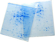
The result of this is a gel with proteins spread out on its surface. These proteins can then be detected by a variety of means, but the most commonly used stains are silver
Silver stain
Silver staining is the use of silver to selectively alter the appearance of the target.-Use in medicine:It is used to stain histologic sections. This kind of staining is important especially to show proteins and DNA. It is used to show both substances inside and outside cells...
and Coomassie Brilliant Blue staining. In this case, a silver colloid is applied to the gel. The silver binds to cysteine groups within the protein. The silver is darkened by exposure to ultra-violet light. The darkness of the silver can be related to the amount of silver and therefore the amount of protein at a given location on the gel. This measurement can only give approximate amounts, but is adequate for most purposes.
Molecules other than proteins can be separated by 2D electrophoresis. In supercoiling assays, coiled DNA
DNA
Deoxyribonucleic acid is a nucleic acid that contains the genetic instructions used in the development and functioning of all known living organisms . The DNA segments that carry this genetic information are called genes, but other DNA sequences have structural purposes, or are involved in...
is separated in the first dimension and denatured by a DNA intercalator (such as ethidium bromide
Ethidium bromide
Ethidium bromide is an intercalating agent commonly used as a fluorescent tag in molecular biology laboratories for techniques such as agarose gel electrophoresis. It is commonly abbreviated as "EtBr", which is also an abbreviation for bromoethane...
or the less carcinogenic chloroquine
Chloroquine
Chloroquine is a 4-aminoquinoline drug used in the treatment or prevention of malaria.-History:Chloroquine , N'--N,N-diethyl-pentane-1,4-diamine, was discovered in 1934 by Hans Andersag and co-workers at the Bayer laboratories who named it "Resochin". It was ignored for a decade because it was...
) in the second. This is comparable to the combination of native PAGE /SDS-PAGE in protein separation.
In summary, 2D gel electrophoresis provides resolution according to two traits, whereof one is most often molecular charge. The investigated molecule need not be protein.
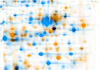
2D gel analysis software

Quantitative proteomics
The aim of quantitative proteomics is to obtain quantitative information about all proteins in a sample. Rather than just providing lists of proteins identified in a certain sample, quantitative proteomics yields information about differences between samples. For example, this approach can be used...
, these tools primarily analyze bio-markers by quantifying individual proteins, and showing the separation between one or more protein "spots" on a scanned image of a 2-DE gel. Additionally, these tools match spots between gels of similar samples to show, for example, proteomic differences between early and advanced stages of an illness. Software packages include Delta2D, ImageMaster, Melanie, PDQuest, Progenesis and REDFIN - among others. While this technology is widely utilized, the intelligence has not been perfected. For example, while PDQuest and Progenesis tend to agree on the quantification and analysis of well-defined well-separated protein spots, they deliver different results and analysis tendencies with less-defined less-separated spots.
Challenges for automatic software-based analysis include:
- incompletely separated (overlapping) spots (less-defined and/or separated)
- weak spots / noise (e.g., "ghost spots")
- running differences between gels (e.g., protein migrates to different positions on different gels)
- unmatched/undetected spots, leading to missing valuesMissing valuesIn statistics, missing data, or missing values, occur when no data value is stored for the variable in the current observation. Missing data are a common occurrence and can have a significant effect on the conclusions that can be drawn from the data....
- mismatched spots
- errors in quantification (several distinct spots may be erroneously detected as a single spot by the software and/or parts of a spot may be excluded from quantification)
- differences in software algorithms and therefore analysis tendencies
Generated picking lists can be used for the automated in-gel digestion
In-gel digestion
The in-gel digestion is part of the sample preparation for the mass spectrometric identification of proteins in course of proteomic analysis. The method was introduced 1992 by Rosenfeld...
of protein spots, and subsequent identification of the proteins by mass spectrometry
Protein mass spectrometry
Protein mass spectrometry refers to the application of mass spectrometry to the study of proteins. Mass spectrometry is an important emerging method for the characterization of proteins. The two primary methods for ionization of whole proteins are electrospray ionization and matrix-assisted laser...
.
For an overview of the current approach for software analysis of 2DE gel images see or.
See also
- Difference gel electrophoresisDifference gel electrophoresis'Difference gel electrophoresis' is a form of gel electrophoresis where up to three different protein samples can be labeled with fluorescent dyes prior to two-dimensional electrophoresis. After the gel electrophoresis, the gel is scanned with the excitation wavelength of each dye one after the...
- QPNC-PAGEQPNC-PAGEQPNC-PAGE, or quantitative preparative native continuous polyacrylamide gel electrophoresis, is a high-resolution technique applied in biochemistry and bioinorganic chemistry to separate proteins by isoelectric point...
- PROTOMAPProtomap (proteomics)PROTOMAP is a recently developed proteomic technology for identifying changes to proteins that manifest in altered migration by one-dimensional SDS-PAGE...
External links
- A 2-D electrophoresis tutorial on the web site of the Parasitology Group at Aberystwyth University
- JVirGel Create virtual 2-D Gels from sequence data.
- fixingproteomics.org Protocols for preparing samples and running 2-D Gels.nnn
- Gel IQ A freely downloadable software tool for assessing the quality of 2D gel image analysis data.