
Inner ear
Encyclopedia
Ear
The ear is the organ that detects sound. It not only receives sound, but also aids in balance and body position. The ear is part of the auditory system....
. In mammal
Mammal
Mammals are members of a class of air-breathing vertebrate animals characterised by the possession of endothermy, hair, three middle ear bones, and mammary glands functional in mothers with young...
s, it consists of the bony labyrinth, a hollow cavity in the temporal bone of the skull with a system of passages comprising two main functional parts:
- The cochleaCochleaThe cochlea is the auditory portion of the inner ear. It is a spiral-shaped cavity in the bony labyrinth, making 2.5 turns around its axis, the modiolus....
, dedicating to hearing; converting sound pressure impulses from the outer ear into electrical impulses which are passed on to the brain via the auditory nerve. - The vestibular systemVestibular systemThe vestibular system, which contributes to balance in most mammals and to the sense of spatial orientation, is the sensory system that provides the leading contribution about movement and sense of balance. Together with the cochlea, a part of the auditory system, it constitutes the labyrinth of...
, dedicated to balanceBalance (ability)In biomechanics, balance is an ability to maintain the center of gravity of a body within the base of support with minimal postural sway. When exercising the ability to balance, one is said to be balancing....
The inner ear is found in all vertebrates, with substantial variations in form and function. The inner ear is innervated by the eighth cranial nerve in all vertebrates.
Embryology
The human inner ear develops during week 4 of embryonic development from the auditory placode, a thickening of the ectodermEctoderm
The "ectoderm" is one of the three primary germ cell layers in the very early embryo. The other two layers are the mesoderm and endoderm , with the ectoderm as the most exterior layer...
which gives rise to the bipolar neurons of the cochlear
Scarpa's ganglion
The vestibular nerve ganglion is the ganglion of the vestibular nerve. It contains the cell bodies of the bipolar primary afferent neurons whose peripheral processes form synaptic contact with hair cells of the vestibular sensory end organs.It is named for Antonio Scarpa.At birth, it is already...
and vestibular ganglions
Spiral ganglion
The spiral ganglion is the group of nerve cells that serve the sense of hearing by sending a representation of sound from the cochlea to the brain...
. As the auditory placode invaginates towards the embryonic mesoderm
Mesoderm
In all bilaterian animals, the mesoderm is one of the three primary germ cell layers in the very early embryo. The other two layers are the ectoderm and endoderm , with the mesoderm as the middle layer between them.The mesoderm forms mesenchyme , mesothelium, non-epithelial blood corpuscles and...
, it forms the auditory vesicle or otocysts.
The auditory vesicle
Auditory vesicle
When the mouth of the auditory pit is closed, and thus a shut sac, the auditory vesicle , is formed; from it the epithelial lining of the membranous labyrinth is derived....
will give rise to the utricluar and saccular components of the membranous labyrinth
Membranous labyrinth
The receptors for the senses of equilibrium and hearing are housed within a collection of fluid filled tubes and chambers known as the membranous labyrinth...
. They contain the sensory hair cells and otolith
Otolith
An otolith, , also called statoconium or otoconium is a structure in the saccule or utricle of the inner ear, specifically in the vestibular labyrinth of vertebrates. The saccule and utricle, in turn, together make the otolith organs. They are sensitive to gravity and linear acceleration...
s of the macula of utricle
Macula of utricle
The portion of the utricle which is lodged in the recess forms a sort of pouch or cul-de-sac, the floor and anterior wall of which are thickened, and form the macula of utricle , which receives the utricular filaments of the vestibulocochlear nerve.The macula of utricle allows a person to perceive...
and of the saccule
Macula of saccule
The saccule is the smaller of the two vestibular sacs; it is globular in form, and lies in the recessus sphæricus near the opening of the scala vestibuli of the cochlea. Its anterior part exhibits an oval thickening, the macula of saccule , to which are distributed the saccular filaments of the...
, respectively, which respond to linear acceleration and the force of gravity. The utricular division of the auditory vesicle also responds to angular acceleration
Angular acceleration
Angular acceleration is the rate of change of angular velocity over time. In SI units, it is measured in radians per second squared , and is usually denoted by the Greek letter alpha .- Mathematical definition :...
, as well as the endolymphatic sac
Endolymphatic sac
From the posterior wall of the saccule a canal, the ductus endolymphaticus, is given off; this duct is joined by the ductus utriculosaccularis, and then passes along the aquaeductus vestibuli and ends in a blind pouch, the endolymphatic sac, on the posterior surface of the petrous portion of the...
and duct
Endolymphatic duct
From the posterior wall of the saccule a canal, the endolymphatic duct, is given off; this duct is joined by the ductus utriculosaccularis, and then passes along the aquaeductus vestibuli and ends in a blind pouch on the posterior surface of the petrous portion of the temporal bone, where it is in...
that connect the saccule and utricle.
Beginning in the fifth week of development, the auditory vesicle also gives rise to the cochlear duct, which contains the spiral organ of Corti
Organ of Corti
The organ of Corti is the organ in the inner ear of mammals that contains auditory sensory cells, or "hair cells."The organ was named after the Italian anatomist Marquis Alfonso Giacomo Gaspare Corti , who conducted microscopic research of the mammaliean auditory system.-Structure and function:The...
and the endolymph
Endolymph
Endolymph is the fluid contained in the membranous labyrinth of the inner ear. It is also called Scarpa's fluid, after Antonio Scarpa.-Composition:...
that accumulates in the membranous labyrinth. The vestibular wall
Reissner's membrane
Reissner's membrane is a membrane inside the cochlea of the inner ear. It separates scala media from scala vestibuli. Together with the basilar membrane it creates a compartment in the cochlea filled with endolymph, which is important for the function of the organ of Corti...
will separate the cochlear duct from the perilymphatic scala vestibuli
Scala vestibuli
Scala vestibuli is a perilymph-filled cavity inside the cochlea of the inner ear that conducts sound vibrations to the scala media.It is separated from the scala media by Reissner's membrane and extends from the vestibule of the ear to the helicotrema where it joins scala tympani.-External links:* ...
, a cavity inside the cochlea. The basilar membrane
Basilar membrane
The basilar membrane within the cochlea of the inner ear is a stiff structural element that separates two liquid-filled tubes that run along the coil of the cochlea, the scala media and the scala tympani .-Function:...
separates the cochlear duct from the scala tympani
Scala tympani
Scala tympani is one of the perilymph-filled cavities in the cochlear labyrinth of the human ear. It is separated from the scala media by the basilar membrane, and it extends from the round window to the helicotrema, where it continues as scala vestibuli....
, a cavity within the cochlear labyrinth. The lateral wall of the cochlear duct is formed by the spiral ligament
Spiral ligament
The periosteum, forming the outer wall of the ductus cochlearis, is greatly thickened and altered in character, and is called the spiral ligament.-External links:* * at IUPUI...
and the stria vascularis
Stria vascularis
The upper portion of the spiral ligament contains numerous capillary loops and small blood vessels, and is termed the stria vascularis. It produces endolymph for the scala media, one of the three fluid-filled compartments of the cochlea...
, which produces the endolymph
Endolymph
Endolymph is the fluid contained in the membranous labyrinth of the inner ear. It is also called Scarpa's fluid, after Antonio Scarpa.-Composition:...
. The hair cells develop from the lateral and medial ridges of the cochlear duct, which together with the tectorial membrane
Tectorial membrane
Tectorial membrane can refer to:*Tectorial membrane *Tectorial membrane...
make up the organ of Corti.
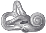
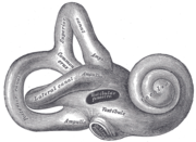
Bony vs. membranous
The bony labyrinth, or osseous labyrinth, is the network of passages with bony walls lined with periosteumPeriosteum
Periosteum is a membrane that lines the outer surface of all bones, except at the joints of long bones. Endosteum lines the inner surface of all bones....
. The membranous labyrinth
Membranous labyrinth
The receptors for the senses of equilibrium and hearing are housed within a collection of fluid filled tubes and chambers known as the membranous labyrinth...
runs inside of the bony labryinth. There is a layer of perilymph
Perilymph
Perilymph is an extracellular fluid located within the cochlea in two of its three compartments: the scala tympani and scala vestibuli. The ionic composition of perilymph is comparable to that of plasma and cerebrospinal fluid...
fluid between them. The three parts of the bony labyrinth are the vestibule of the ear
Vestibule of the ear
-Definition:The vestibule is the central part of the osseous labyrinth, and is situated medial to the tympanic cavity, behind the cochlea, and in front of the semicircular canals.The etymology comes from the Latin vestibulum, literally an entrance hall....
, the semicircular canals, and the cochlea.
Vestibular vs. cochlear
In the middle earMiddle ear
The middle ear is the portion of the ear internal to the eardrum, and external to the oval window of the cochlea. The mammalian middle ear contains three ossicles, which couple vibration of the eardrum into waves in the fluid and membranes of the inner ear. The hollow space of the middle ear has...
, the energy of pressure waves
P-wave
P-waves are a type of elastic wave, also called seismic waves, that can travel through gases , solids and liquids, including the Earth. P-waves are produced by earthquakes and recorded by seismographs...
is translated into mechanical vibrations. The cochlea propagates these mechanical signals as waves in fluid and membranes, and finally transduces them to nerve impulses which then are transmitted to the brain.
The vestibular system is the region of the inner ear where the semicircular canals converge, close to the cochlea. The vestibular system works with the visual system to keep objects in focus when the head is moving. Joint and muscle receptors also are important in maintaining balance. The brain receives, interprets, and processes the information from these systems to control balance.
The vestibular system of the inner ear is responsible for the sensations of balance and motion. It uses the same kinds of fluids and detection cells (hair cells) as the cochlea uses, and sends information to the brain about the attitude, rotation, and linear motion of the head. The type of motion or attitude detected by a hair cell depends on its associated mechanical structures, such as the curved tube of a semicircular canal or the calcium carbonate crystals (otolith
Otolith
An otolith, , also called statoconium or otoconium is a structure in the saccule or utricle of the inner ear, specifically in the vestibular labyrinth of vertebrates. The saccule and utricle, in turn, together make the otolith organs. They are sensitive to gravity and linear acceleration...
) of the saccule
Saccule
The saccule is a bed of sensory cells situated in the inner ear. The saccule translates head movements into neural impulses which the brain can interpret. The saccule is sensitive to linear translations of the head, specifically movements up and down...
and utricle.
Pathology
Interference with or infection of the labyrinth can result in a syndrome of ailments called labyrinthitisLabyrinthitis
Labyrinthitis is an inflammation of the inner ear, and a form of unilateral vestibular dysfunction. It derives its name from the labyrinths that house the vestibular system . Labyrinthitis can cause balance disorders....
. The symptoms of Labyrinthitis include temporary nausea, disorientation, vertigo, and dizziness. Labyrinthitis can be caused by viral infections, bacterial infections, or physical blockage of the inner ear.
Anatomical details
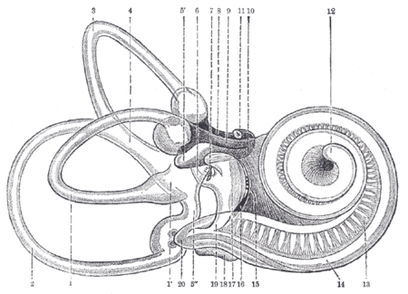
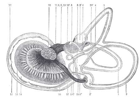
- Lateral semicircular canal; 1’, its ampullaOsseous ampullaeThe bony semicircular canals are three in number, superior, posterior, and lateral, and are situated above and behind the vestibule. They are unequal in length, compressed from side to side, and each describes the greater part of a circle. Each measures about 0.8 mm...
- Posterior canal; 2’, its ampulla
- Superior canal; 3’, its ampulla
- Conjoined limb of superior and posterior canals (sinus utriculi superior)
- Utricle; 5’. Recessus utriculi; 5”. Sinus utriculi posterior
- Ductus endolymphaticus
- Canalis utriculosaccularis
- Nerve to ampulla of superior canal
- Nerve to ampulla of lateral canal
- Nerve to recessus utriculi (in top image, the three branches appear conjoined); 10’. Ending of nerve in recessus utriculi
- Facial nerveFacial nerveThe facial nerve is the seventh of twelve paired cranial nerves. It emerges from the brainstem between the pons and the medulla, and controls the muscles of facial expression, and functions in the conveyance of taste sensations from the anterior two-thirds of the tongue and oral cavity...
- Lagena cochleæ
- Nerve of cochlea within spiral lamina
- Basilar membraneBasilar membraneThe basilar membrane within the cochlea of the inner ear is a stiff structural element that separates two liquid-filled tubes that run along the coil of the cochlea, the scala media and the scala tympani .-Function:...
- Nerve fibers to macula of saccule
- Nerve to ampulla of posterior canal
- SacculeSacculeThe saccule is a bed of sensory cells situated in the inner ear. The saccule translates head movements into neural impulses which the brain can interpret. The saccule is sensitive to linear translations of the head, specifically movements up and down...
- Secondary membrane of tympanum
- Canalis reuniens
- Vestibular end of ductus cochlearis
- Section of the facial and acoustic nerves within internal acoustic meatus (the separation between them is not apparent in the section)
- (No entry)
- Vestibulocochlear nerveVestibulocochlear nerveThe vestibulocochlear nerve is the eighth of twelve cranial nerves, and is responsible for transmitting sound and equilibrium information from the inner ear to the brain...
(auditory or acoustic, cranial nerve VIII)
Non-humans
Birds have an auditory system similar to that of mammals, including a cochlea. Reptiles, amphibians, and fish do not have cochleas but hear with simpler auditory organs or vestibular organs, which generally detect lower-frequency sounds than the cochlea.The cochlear system
In reptileReptile
Reptiles are members of a class of air-breathing, ectothermic vertebrates which are characterized by laying shelled eggs , and having skin covered in scales and/or scutes. They are tetrapods, either having four limbs or being descended from four-limbed ancestors...
s, sound is transmitted to the inner ear by the stapes (stirrup) bone of the middle ear. This is pressed against the oval window
Oval window
The oval window is a membrane-covered opening which leads from the middle ear to the vestibule of the inner ear.Vibrations that come into contact with the tympanic membrane travel through the three ossicles and into the inner ear...
, a membrane-covered opening on the surface of the vestibule. From here, sound waves are conducted through a short perilymphatic duct to a second opening, the round window
Round window
The round window is one of the two openings into the inner ear. It is closed off from the middle ear by the round window membrane, which vibrates with opposite phase to vibrations entering the inner ear through the oval window...
, which equalizes pressure, allowing the incompressible fluid to move freely. Running parallel with the perilymphatic duct is a separate blind-ending duct, the lagena, filled with endolymph
Endolymph
Endolymph is the fluid contained in the membranous labyrinth of the inner ear. It is also called Scarpa's fluid, after Antonio Scarpa.-Composition:...
. The lagena is separated from the perilymphatic duct by a basilar membrane
Basilar membrane
The basilar membrane within the cochlea of the inner ear is a stiff structural element that separates two liquid-filled tubes that run along the coil of the cochlea, the scala media and the scala tympani .-Function:...
, and contains the sensory hair cells that finally translate the vibrations in the fluid into nerve signals. It is attached at one end to the saccule.
In most reptiles the perilymphatic duct and lagena are relatively short, and the sensory cells are confined to a small basilar papilla lying between them. However, in bird
Bird
Birds are feathered, winged, bipedal, endothermic , egg-laying, vertebrate animals. Around 10,000 living species and 188 families makes them the most speciose class of tetrapod vertebrates. They inhabit ecosystems across the globe, from the Arctic to the Antarctic. Extant birds range in size from...
s, mammal
Mammal
Mammals are members of a class of air-breathing vertebrate animals characterised by the possession of endothermy, hair, three middle ear bones, and mammary glands functional in mothers with young...
s, and crocodilians, these structures become much larger and somewhat more complicated. In birds, crocodilians, and monotreme
Monotreme
Monotremes are mammals that lay eggs instead of giving birth to live young like marsupials and placental mammals...
s, the ducts are simply extended, together forming an elongated, more or less straight, tube. The endolymphatic duct is wrapped in a simple loop around the lagena, with the basilar membrane lying along one side. The first half of the duct is now referred to as the scala vestibuli
Scala vestibuli
Scala vestibuli is a perilymph-filled cavity inside the cochlea of the inner ear that conducts sound vibrations to the scala media.It is separated from the scala media by Reissner's membrane and extends from the vestibule of the ear to the helicotrema where it joins scala tympani.-External links:* ...
, while the second half, which includes the basilar membrane, is called the scala tympani
Scala tympani
Scala tympani is one of the perilymph-filled cavities in the cochlear labyrinth of the human ear. It is separated from the scala media by the basilar membrane, and it extends from the round window to the helicotrema, where it continues as scala vestibuli....
. As a result of this increase in length, the basilar membrane and papilla are both extended, with the latter developing into the organ of Corti
Organ of Corti
The organ of Corti is the organ in the inner ear of mammals that contains auditory sensory cells, or "hair cells."The organ was named after the Italian anatomist Marquis Alfonso Giacomo Gaspare Corti , who conducted microscopic research of the mammaliean auditory system.-Structure and function:The...
, while the lagena is now called the cochlear duct. All of these structures together constitute the cochlea.
In mammals (other than monotremes), the cochlea is extended still further, becoming a coiled structure in order to accommodate its length within the head. The organ of Corti also has a more complex structure in mammals than it does in other amniote
Amniote
The amniotes are a group of tetrapods that have a terrestrially adapted egg. They include synapsids and sauropsids , as well as their fossil ancestors. Amniote embryos, whether laid as eggs or carried by the female, are protected and aided by several extensive membranes...
s.
The arrangement of the inner ear in living amphibian
Amphibian
Amphibians , are a class of vertebrate animals including animals such as toads, frogs, caecilians, and salamanders. They are characterized as non-amniote ectothermic tetrapods...
s is, in most respects, similar to that of reptiles. However, they often lack a basilar papilla, having instead an entirely separate set of sensory cells at the upper edge of the saccule, referred to as the papilla amphibiorum, which appear to have the same function.
Although many fish are capable of hearing, the lagena is, at best, a short diverticulum of the saccule, and appears to have no role in sensation of sound. Various clusters of hair cells within the inner ear may instead be responsible; for example, bony fish
Osteichthyes
Osteichthyes , also called bony fish, are a taxonomic group of fish that have bony, as opposed to cartilaginous, skeletons. The vast majority of fish are osteichthyes, which is an extremely diverse and abundant group consisting of over 29,000 species...
contain a sensory cluster called the macula neglecta in the utricle that may have this function. Although fish have neither an outer nor a middle ear, sound may still be transmitted to the inner ear through the bones of the skull, or by the swim bladder, parts of which often lie close by in the body.
The vestibular system
By comparison with the cochlear system, the vestibular system varies relatively little between the various groups of jawed vertebrates. The central part of the system consists of two chambers, the saccule and utricle, each of which includes one or two small clusters of sensory hair cells. All jawed vertebrates also possess three semicircular canals arising from the utricle, each with an ampullaAmpulla
An ampulla was, in Ancient Rome, a "small nearly globular flask or bottle, with two handles" . The word is used of these in archaeology, and of later flasks, often handle-less and much flatter, for holy water or holy oil in the Middle Ages....
containing sensory cells at one end.
An endolymphatic duct
Endolymphatic duct
From the posterior wall of the saccule a canal, the endolymphatic duct, is given off; this duct is joined by the ductus utriculosaccularis, and then passes along the aquaeductus vestibuli and ends in a blind pouch on the posterior surface of the petrous portion of the temporal bone, where it is in...
runs from the saccule up through the head, and ending close to the brain. In cartilaginous fish, this duct actually opens onto the top of the head, and in some teleosts, it is simply blind-ending. In all other species, however, it ends in an endolymphatic sac
Endolymphatic sac
From the posterior wall of the saccule a canal, the ductus endolymphaticus, is given off; this duct is joined by the ductus utriculosaccularis, and then passes along the aquaeductus vestibuli and ends in a blind pouch, the endolymphatic sac, on the posterior surface of the petrous portion of the...
. In many reptiles, fish, and amphibians this sac may reach considerable size. In amphibians the sacs from either side may fuse into a single structure, which often extends down the length of the body, parallel with the spinal canal
Spinal canal
The spinal canal is the space in vertebrae through which the spinal cord passes. It is a process of the dorsal human body cavity. This canal is enclosed within the vertebral foramen of the vertebrae...
.
The primitive lamprey
Lamprey
Lampreys are a family of jawless fish, whose adults are characterized by a toothed, funnel-like sucking mouth. Translated from an admixture of Latin and Greek, lamprey means stone lickers...
s and hagfish
Hagfish
Hagfish, the clade Myxini , are eel-shaped slime-producing marine animals . They are the only living animals that have a skull but not a vertebral column. Along with lampreys, hagfish are jawless and are living fossils whose next nearest relatives include all vertebrates...
, however, have a simpler system. The inner ear in these species consists of a single vestibular chamber, although in lampreys, this is associated with a series of sacs lined by cilia. Lampreys have only two semicircular canals, with the horizontal canal being absent, while hagfish have only a single, vertical, canal.