Excitatory postsynaptic potential
Encyclopedia


Neuroscience
Neuroscience is the scientific study of the nervous system. Traditionally, neuroscience has been seen as a branch of biology. However, it is currently an interdisciplinary science that collaborates with other fields such as chemistry, computer science, engineering, linguistics, mathematics,...
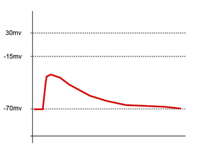
, an excitatory postsynaptic potential (EPSP) is a temporary depolarization of postsynaptic membrane potential
Membrane potential
Membrane potential is the difference in electrical potential between the interior and exterior of a biological cell. All animal cells are surrounded by a plasma membrane composed of a lipid bilayer with a variety of types of proteins embedded in it...
caused by the flow of positively charged ion
Ion
An ion is an atom or molecule in which the total number of electrons is not equal to the total number of protons, giving it a net positive or negative electrical charge. The name was given by physicist Michael Faraday for the substances that allow a current to pass between electrodes in a...
s into the postsynaptic cell as a result of opening of ligand-sensitive channels. They are the opposite of inhibitory postsynaptic potential
Inhibitory postsynaptic potential
An inhibitory postsynaptic potential is a synaptic potential that decreases the chance that a future action potential will occur in a postsynaptic neuron or α-motoneuron...
s (IPSPs), which usually result from the flow of negative ions into the cell or positive ions out of the cell. A postsynaptic potential
Postsynaptic potential
Postsynaptic potentials are changes in the membrane potential of the postsynaptic terminal of a chemical synapse. Postsynaptic potentials are graded potentials, and should not be confused with action potentials although their function is to initiate or inhibit action potentials...
is defined as excitatory if it makes it easier for the neuron to fire an action potential
Action potential
In physiology, an action potential is a short-lasting event in which the electrical membrane potential of a cell rapidly rises and falls, following a consistent trajectory. Action potentials occur in several types of animal cells, called excitable cells, which include neurons, muscle cells, and...
. EPSPs can also result from a decrease in outgoing positive charges, while IPSPs are sometimes caused by an increase in positive charge outflow. The flow of ions that causes an EPSP is an excitatory postsynaptic current (EPSC).
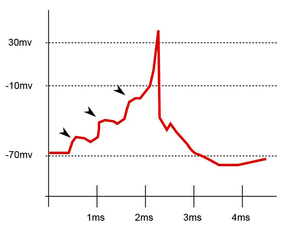
EPSPs, like IPSPs, are graded (i.e. they have an additive effect). When multiple EPSPs occur on a single patch of postsynaptic membrane, their combined effect is the sum of the individual EPSPs. Larger EPSPs result in greater membrane depolarization and thus increase the likelihood that the postsynaptic cell reaches the threshold for firing an action potential
Action potential
In physiology, an action potential is a short-lasting event in which the electrical membrane potential of a cell rapidly rises and falls, following a consistent trajectory. Action potentials occur in several types of animal cells, called excitable cells, which include neurons, muscle cells, and...
.
Overview
EPSPs in living cells are caused chemically. When an active presynaptic cell releases neurotransmitterNeurotransmitter
Neurotransmitters are endogenous chemicals that transmit signals from a neuron to a target cell across a synapse. Neurotransmitters are packaged into synaptic vesicles clustered beneath the membrane on the presynaptic side of a synapse, and are released into the synaptic cleft, where they bind to...
s into the synapse, some of them bind to receptors
Neurotransmitter receptor
A Neurotransmitter receptor is a membrane receptor protein that is activated by a Neurotransmitter. A membrane protein interacts with the lipid bilayer that encloses the cell and a membrane receptor protein interacts with a chemical in the cells external environment, which binds to the cell...
on the postsynaptic cell. Many of these receptors contain an ion channel
Ion channel
Ion channels are pore-forming proteins that help establish and control the small voltage gradient across the plasma membrane of cells by allowing the flow of ions down their electrochemical gradient. They are present in the membranes that surround all biological cells...
capable of passing positively charged ions either into or out of the cell (such receptors are called ionotropic receptors). At excitatory synapses, the ion channel typically allows sodium into the cell, generating an excitatory postsynaptic current. This depolarizing current causes an increase in membrane potential, the EPSP.
Excitatory molecules
The neurotransmitter most often associated with EPSPs is the amino acidAmino acid
Amino acids are molecules containing an amine group, a carboxylic acid group and a side-chain that varies between different amino acids. The key elements of an amino acid are carbon, hydrogen, oxygen, and nitrogen...
glutamate, and is the main excitatory neurotransmitter in the central nervous system
Central nervous system
The central nervous system is the part of the nervous system that integrates the information that it receives from, and coordinates the activity of, all parts of the bodies of bilaterian animals—that is, all multicellular animals except sponges and radially symmetric animals such as jellyfish...
of vertebrates. Its ubiquity at excitatory synapses has led to it being called the excitatory neurotransmitter. In some invertebrates, glutamate is the main excitatory transmitter at the neuromuscular junction
Neuromuscular junction
A neuromuscular junction is the synapse or junction of the axon terminal of a motor neuron with the motor end plate, the highly-excitable region of muscle fiber plasma membrane responsible for initiation of action potentials across the muscle's surface, ultimately causing the muscle to contract...
. In the neuromuscular junction
Neuromuscular junction
A neuromuscular junction is the synapse or junction of the axon terminal of a motor neuron with the motor end plate, the highly-excitable region of muscle fiber plasma membrane responsible for initiation of action potentials across the muscle's surface, ultimately causing the muscle to contract...
of vertebrates, EPP (end-plate potential
End-plate potential
End plate potentials are the depolarizations of skeletal muscle fibers caused by neurotransmitters binding to the postsynaptic membrane in the neuromuscular junction. They are called "end plates" because the postsynaptic terminals of muscle fibers have a large, saucer-like appearance...
s) are mediated by the neurotransmitter acetylcholine
Acetylcholine
The chemical compound acetylcholine is a neurotransmitter in both the peripheral nervous system and central nervous system in many organisms including humans...
, which is also the main transmitter in an invertebrates´ central nervous system.
At the same time, GABA is the most common neurotransmitter associated with IPSPs in the brain.
However, classifying neurotransmitters as such is technically incorrect, as there are several other synaptic factors that help determine a neurotransmitter's excitatory or inhibitory effects.
Miniature EPSPs
The release of neurotransmitter vesiclesSynaptic vesicle
In a neuron, synaptic vesicles store various neurotransmitters that are released at the synapse. The release is regulated by a voltage-dependent calcium channel. Vesicles are essential for propagating nerve impulses between neurons and are constantly recreated by the cell...
from the presynaptic cell is probabilistic. In fact, even without stimulation of the presynaptic cell, a single vesicle will occasionally be released into the synapse, generating miniature EPSPs
(mEPSPs). Bernard Katz
Bernard Katz
Sir Bernard Katz, FRS was a German-born biophysicist, noted for his work on nerve biochemistry. He shared the Nobel Prize in physiology or medicine in 1970 with Julius Axelrod and Ulf von Euler...
pioneered the study of these mEPSPs at the neuromuscular junction
Neuromuscular junction
A neuromuscular junction is the synapse or junction of the axon terminal of a motor neuron with the motor end plate, the highly-excitable region of muscle fiber plasma membrane responsible for initiation of action potentials across the muscle's surface, ultimately causing the muscle to contract...
(often called miniature end-plate potentials) in 1951, revealing the quantal nature of synaptic transmission. Quantal size can then be defined as the synaptic response to the release of neurotransmitter from a single vesicle, while quantal content is the number of effective vesicles released in response to a nerve impulse.
Field EPSPs
EPSPs are usually recorded using intracellular electrodes. The extracellular signal from a single neuron is extremely small and thus next to impossible to record in the human brain. However, in some areas of the brain, such as the hippocampusHippocampus
The hippocampus is a major component of the brains of humans and other vertebrates. It belongs to the limbic system and plays important roles in the consolidation of information from short-term memory to long-term memory and spatial navigation. Humans and other mammals have two hippocampi, one in...
, neurons are arranged in such a way that they all receive synaptic inputs in the same area. Because these neurons are in the same orientation, the extracellular signals from synaptic excitation don't cancel out, but rather add up to give a signal that can easily be recorded with a field electrode. This extracellular signal recorded from a population of neurons is the field potential. In studies of hippocampal LTP
Long-term potentiation
In neuroscience, long-term potentiation is a long-lasting enhancement in signal transmission between two neurons that results from stimulating them synchronously. It is one of several phenomena underlying synaptic plasticity, the ability of chemical synapses to change their strength...
, figures are often given showing the field EPSP (fEPSP) in stratum radiatum of CA1 in response to Schaffer collateral stimulation. This is the signal seen by an extracellular electrode placed in the layer of apical dendrites of CA1 pyramidal neurons. The Schaffer collaterals make excitatory synapses onto these dendrites, and so when they are activated, there is a current sink in stratum radiatum: the field EPSP. The voltage deflection recorded during a field EPSP is negative-going, while an intracellularly recorded EPSP is positive-going. This difference is due to the relative flow of ions (primarily the sodium ion) into the cell, which, in the case of the field EPSP is away from the electrode, while for an intracellular EPSPs it is towards the electrode. After a field EPSP, the extracellular electrode may record another change in electrical potential named the population spike
Population spike
In neuroscience, a population spike is the shift in electrical potential as a consequence of the movement of ions involved in the generation and propagation of action potentials...
which corresponds to the population of cells firing action potentials (spiking). In other regions than CA1 of the hippocampus, the field EPSP may be far more complex and harder to interpret as the source and sinks are far less defined. In regions such as the striatum
Striatum
The striatum, also known as the neostriatum or striate nucleus, is a subcortical part of the forebrain. It is the major input station of the basal ganglia system. The striatum, in turn, gets input from the cerebral cortex...
neurotransmitters such as dopamine
Dopamine
Dopamine is a catecholamine neurotransmitter present in a wide variety of animals, including both vertebrates and invertebrates. In the brain, this substituted phenethylamine functions as a neurotransmitter, activating the five known types of dopamine receptors—D1, D2, D3, D4, and D5—and their...
, acetylcholine
Acetylcholine
The chemical compound acetylcholine is a neurotransmitter in both the peripheral nervous system and central nervous system in many organisms including humans...
, GABA
Gabâ
Gabâ or gabaa, for the people in many parts of the Philippines), is the concept of a non-human and non-divine, imminent retribution. A sort of negative karma, it is generally seen as an evil effect on a person because of their wrongdoings or transgressions...
and others may also be released and further complicate the interpretation.
See also
- Inhibitory postsynaptic potentialInhibitory postsynaptic potentialAn inhibitory postsynaptic potential is a synaptic potential that decreases the chance that a future action potential will occur in a postsynaptic neuron or α-motoneuron...
(IPSP) - Postsynaptic potentialPostsynaptic potentialPostsynaptic potentials are changes in the membrane potential of the postsynaptic terminal of a chemical synapse. Postsynaptic potentials are graded potentials, and should not be confused with action potentials although their function is to initiate or inhibit action potentials...
- GABAGabâGabâ or gabaa, for the people in many parts of the Philippines), is the concept of a non-human and non-divine, imminent retribution. A sort of negative karma, it is generally seen as an evil effect on a person because of their wrongdoings or transgressions...
- GlycineGlycineGlycine is an organic compound with the formula NH2CH2COOH. Having a hydrogen substituent as its 'side chain', glycine is the smallest of the 20 amino acids commonly found in proteins. Its codons are GGU, GGC, GGA, GGG cf. the genetic code.Glycine is a colourless, sweet-tasting crystalline solid...

