Electrogastrogram
Encyclopedia
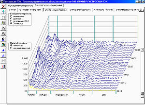
An electrogastrogram is a graphic produced by an electrogastrograph, which records the electrical signals that travel through the stomach
muscles and control the muscles' contractions. An electrogastroenterogram (or gastroenterogram) is a similar procedure, which writes down electric signals not only from the stomach, but also from intestines.
These names are made of different parts: electro, because it is related to electrical activity, gastro, Greek
for stomach, entero, Greek for intestines, gram, a Greek root meaning "to write".
An electrogastrogram and a gastroenterogram are similar in principle to an electrocardiogram
(ECG) in that sensors on the skin detect electrical signals indicative of muscular activity within. Where the electrocardiogram detects muscular activity in various regions of the heart, the electrogastrogram detects the wave-like contractions of the stomach (peristalsis
).
Walter C. Alvarez
pioneered early studies of electrogastrography in 1921-22.
of GI tract is results from coordinated contractions of smooth muscle
, which in turn derive from two basic patterns of electrical activity across the membranes
of smooth muscle cells — slow waves
and action potential
s. Slow waves are initiated by pacemakers — the interstitial cells of Cajal
(ICC). Slow wave frequency varies in the different organs of the GI tract and is characteristic for that organ. They set the maximum frequency at which the muscle can contract:
The electrical activity of the GI tract can be subdivided into two categories: electrical control activity (ECA) and electrical response activity (ERA). ECA is characterized by regularly recurring electrical potentials, originating in the gastric pacemaker located in the body of stomach
.
The slow waves are not a direct reason of peristalsis of a GI tract, but a correlation between deviations of slow waves from norm and motility abnormalities however is proved.
Several EGG signals are recorded from various standardized positions on the abdominal wall and to select the one with the highest amplitude for further analysis. For this purpose usually use three or more Ag-AgCl
electrodes. Recordings are made both fasting (usually 30 minutes) and after a meal (usually 60 minutes) with the patient lying quietly. Deviations from the normal frequency may be referred to as bradygastria or tachygastria.
In normal individuals the power of the electrical signals increases after the meal. In patients with abnormalities of stomach motility, the rhythm often is irregular or there is no postprandial increase in electrical power.
A bradygastria is defined, how decreased rate of electrical activity in the stomach, as less than 2 cycles per minute for at least 1 minute.
A tachygastria is defined, how increased rate of electrical activity in the stomach, as more than 4 cycles per minute for at least 1 minute.
A bradygastria and tachygastria may be associated with nausea
, gastroparesis
, irritable bowel syndrome
, and functional dyspepsia.
(CPT) and Healthcare Common Procedure Coding System
(HCPCS) codes (maintained by the American Medical Association
) for cutaneous electrogastrography:
EGEG electrodes are as much as possible removed from a stomach and an intestines — usaually three electrodes are placed on the extremities. It allows to receive stabler and comparable results.
 An electrogastroenterography analysis program calculate
where S(n) — spectral components in the rank from sti to fini (defined by received investigated range of this organ of GI tract) by Discrete Fourier transform
of the electric signal from GI tract.
EGEG parametres for normal patients:
and functional dyspepsia.
Stomach
The stomach is a muscular, hollow, dilated part of the alimentary canal which functions as an important organ of the digestive tract in some animals, including vertebrates, echinoderms, insects , and molluscs. It is involved in the second phase of digestion, following mastication .The stomach is...
muscles and control the muscles' contractions. An electrogastroenterogram (or gastroenterogram) is a similar procedure, which writes down electric signals not only from the stomach, but also from intestines.
These names are made of different parts: electro, because it is related to electrical activity, gastro, Greek
Greek language
Greek is an independent branch of the Indo-European family of languages. Native to the southern Balkans, it has the longest documented history of any Indo-European language, spanning 34 centuries of written records. Its writing system has been the Greek alphabet for the majority of its history;...
for stomach, entero, Greek for intestines, gram, a Greek root meaning "to write".
An electrogastrogram and a gastroenterogram are similar in principle to an electrocardiogram
Electrocardiogram
Electrocardiography is a transthoracic interpretation of the electrical activity of the heart over a period of time, as detected by electrodes attached to the outer surface of the skin and recorded by a device external to the body...
(ECG) in that sensors on the skin detect electrical signals indicative of muscular activity within. Where the electrocardiogram detects muscular activity in various regions of the heart, the electrogastrogram detects the wave-like contractions of the stomach (peristalsis
Peristalsis
Peristalsis is a radially symmetrical contraction and relaxation of muscles which propagates in a wave down the muscular tube, in an anterograde fashion. In humans, peristalsis is found in the contraction of smooth muscles to propel contents through the digestive tract. Earthworms use a similar...
).
Walter C. Alvarez
Walter C. Alvarez
Walter Clement Alvarez was an American doctor of Spanish descent. He authored several dozen books on medicine, and wrote Introductions and Forewords for many others....
pioneered early studies of electrogastrography in 1921-22.
Physiological basis of electrogastroenterography
MotilityMotility
Motility is a biological term which refers to the ability to move spontaneously and actively, consuming energy in the process. Most animals are motile but the term applies to single-celled and simple multicellular organisms, as well as to some mechanisms of fluid flow in multicellular organs, in...
of GI tract is results from coordinated contractions of smooth muscle
Smooth muscle
Smooth muscle is an involuntary non-striated muscle. It is divided into two sub-groups; the single-unit and multiunit smooth muscle. Within single-unit smooth muscle tissues, the autonomic nervous system innervates a single cell within a sheet or bundle and the action potential is propagated by...
, which in turn derive from two basic patterns of electrical activity across the membranes
Cell membrane
The cell membrane or plasma membrane is a biological membrane that separates the interior of all cells from the outside environment. The cell membrane is selectively permeable to ions and organic molecules and controls the movement of substances in and out of cells. It basically protects the cell...
of smooth muscle cells — slow waves
Slow wave potential
In physiology, a slow-wave potential is a membrane potential that cycles between depolarizations and repolarizations. Slow wave potentials are generated by myocytes. Due to temporal summation, a slow-wave potential will periodically reach threshold and generate an action potential. This in turn...
and action potential
Action potential
In physiology, an action potential is a short-lasting event in which the electrical membrane potential of a cell rapidly rises and falls, following a consistent trajectory. Action potentials occur in several types of animal cells, called excitable cells, which include neurons, muscle cells, and...
s. Slow waves are initiated by pacemakers — the interstitial cells of Cajal
Interstitial cell of Cajal
The Interstitial cell of Cajal is a type of interstitial cell found in the gastrointestinal tract that serves as a pacemaker which creates the basal electrical rhythm leading to contraction of the smooth muscle ....
(ICC). Slow wave frequency varies in the different organs of the GI tract and is characteristic for that organ. They set the maximum frequency at which the muscle can contract:
- stomach — about 3 waves in a minute,
- duodenumDuodenumThe duodenum is the first section of the small intestine in most higher vertebrates, including mammals, reptiles, and birds. In fish, the divisions of the small intestine are not as clear and the terms anterior intestine or proximal intestine may be used instead of duodenum...
— about 12 waves in a minute, - ileumIleumThe ileum is the final section of the small intestine in most higher vertebrates, including mammals, reptiles, and birds. In fish, the divisions of the small intestine are not as clear and the terms posterior intestine or distal intestine may be used instead of ileum.The ileum follows the duodenum...
— about 8 waves in a minute, - rectumRectumThe rectum is the final straight portion of the large intestine in some mammals, and the gut in others, terminating in the anus. The human rectum is about 12 cm long...
— about 17 waves in a minute. - jejunumJejunumThe jejunum is the middle section of the small intestine in most higher vertebrates, including mammals, reptiles, and birds. In fish, the divisions of the small intestine are not as clear and the terms middle intestine or mid-gut may be used instead of jejunum.The jejunum lies between the duodenum...
— about 11 waves in a minute.
The electrical activity of the GI tract can be subdivided into two categories: electrical control activity (ECA) and electrical response activity (ERA). ECA is characterized by regularly recurring electrical potentials, originating in the gastric pacemaker located in the body of stomach
Body of stomach
The Body of the Stomach often just called the body or corpus is an anatomical region of the stomach in humans....
.
The slow waves are not a direct reason of peristalsis of a GI tract, but a correlation between deviations of slow waves from norm and motility abnormalities however is proved.
Cutaneous electrogastrography
Electrogastrogram can be made from the gastrointestinal mucosa, serosa, or skin surface. The cutaneous electrogastrography and provides an indirect representation of the electrical activity but it is much easier and therefore cutaneous electrogastrography has been used most frequently.Several EGG signals are recorded from various standardized positions on the abdominal wall and to select the one with the highest amplitude for further analysis. For this purpose usually use three or more Ag-AgCl
Silver chloride electrode
A silver chloride electrode is a type of reference electrode, commonly used in electrochemical measurements. For example, it is usually the internal reference electrode in pH meters...
electrodes. Recordings are made both fasting (usually 30 minutes) and after a meal (usually 60 minutes) with the patient lying quietly. Deviations from the normal frequency may be referred to as bradygastria or tachygastria.
In normal individuals the power of the electrical signals increases after the meal. In patients with abnormalities of stomach motility, the rhythm often is irregular or there is no postprandial increase in electrical power.
Bradygastria, normogastria and tachygastria
Terms bradygastria and tachygastria are used at the description of deviations of frequency of an electric signal from slow waves are initiated by pacemaker in the stomach from normal frequency of 3 cycles per minute.A bradygastria is defined, how decreased rate of electrical activity in the stomach, as less than 2 cycles per minute for at least 1 minute.
A tachygastria is defined, how increased rate of electrical activity in the stomach, as more than 4 cycles per minute for at least 1 minute.
A bradygastria and tachygastria may be associated with nausea
Nausea
Nausea , is a sensation of unease and discomfort in the upper stomach with an involuntary urge to vomit. It often, but not always, precedes vomiting...
, gastroparesis
Gastroparesis
Gastroparesis, also called delayed gastric emptying, is a medical condition consisting of a paresis of the stomach, resulting in food remaining in the stomach for a longer period of time than normal. Normally, the stomach contracts to move food down into the small intestine for digestion. The...
, irritable bowel syndrome
Irritable bowel syndrome
Irritable bowel syndrome is a diagnosis of exclusion. It is a functional bowel disorder characterized by chronic abdominal pain, discomfort, bloating, and alteration of bowel habits in the absence of any detectable organic cause. In some cases, the symptoms are relieved by bowel movements...
, and functional dyspepsia.
CPT and HCPCS codes for electrogastrography
There are following Current Procedural TerminologyCurrent Procedural Terminology
The Current Procedural Terminology code set is maintained by the American Medical Association through the CPT Editorial Panel. The CPT code set describes medical, surgical, and diagnostic services and is designed to communicate uniform information about medical services and procedures among...
(CPT) and Healthcare Common Procedure Coding System
Healthcare Common Procedure Coding System
The Healthcare Common Procedure Coding System is a set of health care procedure codes based on the American Medical Association's Current Procedural Terminology .-History:...
(HCPCS) codes (maintained by the American Medical Association
American Medical Association
The American Medical Association , founded in 1847 and incorporated in 1897, is the largest association of medical doctors and medical students in the United States.-Scope and operations:...
) for cutaneous electrogastrography:
| CPT/HCPCS-code | Procedure |
|---|---|
| 91132 | Electrogastrography, diagnostic, transcutaneous |
| 91133 | Electrogastrography, diagnostic, transcutaneous; with provocative testing |
Electrogastroenterography
An electrogastroenterography (EGEG) is based that different organs of a GI tract give different frequency slow wave.| Organ of gastrointestinal tract | Investigated range (Hz Hertz The hertz is the SI unit of frequency defined as the number of cycles per second of a periodic phenomenon. One of its most common uses is the description of the sine wave, particularly those used in radio and audio applications.... ) |
Frequency number (i) |
|---|---|---|
| Large intestine Large intestine The large intestine is the third-to-last part of the digestive system — — in vertebrate animals. Its function is to absorb water from the remaining indigestible food matter, and then to pass useless waste material from the body... |
0.01 – 0.03 | 5 |
| Stomach Stomach The stomach is a muscular, hollow, dilated part of the alimentary canal which functions as an important organ of the digestive tract in some animals, including vertebrates, echinoderms, insects , and molluscs. It is involved in the second phase of digestion, following mastication .The stomach is... |
0.03 – 0.07 | 1 |
| Ileum Ileum The ileum is the final section of the small intestine in most higher vertebrates, including mammals, reptiles, and birds. In fish, the divisions of the small intestine are not as clear and the terms posterior intestine or distal intestine may be used instead of ileum.The ileum follows the duodenum... |
0.07 – 0.13 | 4 |
| Jejunum Jejunum The jejunum is the middle section of the small intestine in most higher vertebrates, including mammals, reptiles, and birds. In fish, the divisions of the small intestine are not as clear and the terms middle intestine or mid-gut may be used instead of jejunum.The jejunum lies between the duodenum... |
0.13 – 0.18 | 3 |
| Duodenum Duodenum The duodenum is the first section of the small intestine in most higher vertebrates, including mammals, reptiles, and birds. In fish, the divisions of the small intestine are not as clear and the terms anterior intestine or proximal intestine may be used instead of duodenum... |
0.18 – 0.25 | 2 |
EGEG electrodes are as much as possible removed from a stomach and an intestines — usaually three electrodes are placed on the extremities. It allows to receive stabler and comparable results.
The computer analysis of electrogastroenterograms

- P(i) — capacities of an electric signal separately from each of organ of GI tract in corresponding range of frequencies:
where S(n) — spectral components in the rank from sti to fini (defined by received investigated range of this organ of GI tract) by Discrete Fourier transform
Discrete Fourier transform
In mathematics, the discrete Fourier transform is a specific kind of discrete transform, used in Fourier analysis. It transforms one function into another, which is called the frequency domain representation, or simply the DFT, of the original function...
of the electric signal from GI tract.
- PS — the general (total) capacity of an electric signal from five parts of GI tract:
-

- P(i)/PS — the relative electric activity.
- Kritm(i) — rhythm factor
-

EGEG parametres for normal patients:
| Organ of gastrointestinal tract | Electric activity P(i)/PS | Rhythm factor Kritm(i) | P(i)/P(i+1) |
|---|---|---|---|
| Stomach | 22.4±11.2 | 4.85±2.1 | 10.4±5.7 |
| Duodenum | 2.1±1.2 | 0.9±0.5 | 0.6±0.3 |
| Jejunum | 3.35±1.65 | 3.43±1.5 | 0.4±0.2 |
| Ileum | 8.08±4.01 | 4.99±2.5 | 0.13±0.08 |
| Large intestine | 64.04±32.01 | 22.85±9.8 | — |
Not solved problems
There are some lacks limiting an electrogastroenterography use in practice:- an absence of a standard technique of performance peripheral electrogastroenterography,
- an absence of a standard norms of electrophysiological parametres of bioelectric activity GT tract,
- an impossibility of an estimation of change of motility abnormalities during the concrete moments of time on local sites of GI tract.
Other updatings of electrogastrography
- 24-hour electrogastrography and electrogastroenterography.
- The joint electrogastroenterography with 24-hours pH-metry.
- Wavelet analysis of electrogastroenterogram.
- TelemetryTelemetryTelemetry is a technology that allows measurements to be made at a distance, usually via radio wave transmission and reception of the information. The word is derived from Greek roots: tele = remote, and metron = measure...
capsule for the EGG monitoring in a stomach and an intestines.
Clinical applications of electrogastrography and gastroenterography
Electrogastrography or gastroenterography used when a patient is suspected of having a motility disorder, which can be shown, as recurrent nausea and vomiting, signs that the stomach is not emptying food normally. The clinical use of electrogastrography has been most widely evaluated in patients with gastroparesisGastroparesis
Gastroparesis, also called delayed gastric emptying, is a medical condition consisting of a paresis of the stomach, resulting in food remaining in the stomach for a longer period of time than normal. Normally, the stomach contracts to move food down into the small intestine for digestion. The...
and functional dyspepsia.


