

Carpometacarpal joint
Encyclopedia
The carpometacarpal joints are five joint
s in the wrist
that articulates the distal row of carpal bones
and the proximal bases of the five metacarpal bones.
The CMC of the thumb or the first CMC differs significantly from the other four CMCs and is therefore described separately.
to the first metacarpal bone, plays an irreplaceable role in the normal functioning of the thumb. The most important joint connecting the wrist to the metacarpus, osteoarthritis of the TMC is a severely disabling condition; up to twenty times more common among old women than in average.
Pronation-supination of the first metacarpal is especially important for the pulp-to-pulp pinch (i.e. "true opposition").
The movements of the first CMC is limited by the shape of the joint, by the capsulo-ligamentous complex surrounding the joint, and by the balance among involved muscles. If the first metacarpal fails to sit well 'on the saddle', for example because of hypoplasia
, the first CMC joint tends to be subluxated
(i.e. slightly displaced) towards the radius
.
The capsule is sufficiently slack to allow a wide range of movements and a distraction of roughly 3 mm, while reinforcing ligaments and tendons give stability to the joint. It is slightly thicker on its dorsal side than on the other.
The first carpometacarpal joint is a frequent site of osteoarthritis
in postmenopausal women.
Anterior oblique ligament (AOL): A strong, thick, and extracapsular ligament originating on the palmar tubercle of the trapezium to be inserted on the palmar tubercle of the first metacarpal. It is taut in abduction, extension, and pronation, and has been reported to have an important retaining function and to be elongated or absent in CMC joint arthritis.
Ulnar collateral ligament (UCL): The second extracapsular ligament, the UCL is located ulnarly to the AOL. It has its origin on the flexor retinaculum
and is inserted on the ulnopalmar tubercle of the first metacarpal. It is taut in abduction, extension, and pronation, and often found elongated in connection to CMC joint arthritis. The importance ascribed to the UCL varies considerably among researchers.
First intermetacarpal ligament (IML): Connecting the bases of the second and first metacarpals, this ligament inserts onto the ulnopalmar tubercle of the first metacarpal where its fibers intermingle with those of the UCL. It is taut in abduction, opposition, and supination. It has been reported to be the most important restraining structure of the first CMC joint by several researchers, while some consider it to weak to be able to stabilize the joint by itself, but that it together with the UCL represent an important restraining structure.
Posterior oblique ligment (POL): An intracapsular ligament stretching from the dorsoulnar side of the trapezium to the ulno-palmar tubercle of the first metacarpal. Not considered an important ligament to the first CMC joint, it tightens during forced adduction and radial abduction.
Dorsoradial ligament (DRL): Like the previous ligament, the DRL is not considered important to the first CMC. It connects the dorsal sides of the trapezium and the first metacarpal.
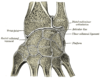
Early, anatomically correct drawings of the ligaments of the first carpometacarpal joints where produced by .
Range of motion
for the first CMC is 53° of flexion/extension, 42° of abduction/adduction, and 17° of rotation
Planes and axes of movements
The thumb's MP and CMC joints abduct and adduct in a plane perpendicular to the palm, a movement also referred to as "palmar abduction." The same joints flex and extend in a plane parallel to the palm, also referred to as "radial abduction," because the thumb moves toward the hand's radial side. Abduction and adduction occur around an antero-posterior axis, while flexion and extension occur around a lateral axis.
For ease of orientation, the thumbnail can be considered as resting in the thumbs frontal plane. Abduction and adduction of the first CMC (and MP) joint(s) occur in this plane; flexion and extension of the first CMC, MP, and IP joints occur in a plane that is perpendicular to the thumbnail. This remains true regardless of how the first metacarpal bone is being rotated during opposition and reposition.
Among themselves, the four ulnar metacarpals also articulates with their neighbours at the intermetacarpal articulations
.
and the volar or palmar carpometacarpal ligaments
.
The interosseous ligaments consist of short, thick fibers, and are limited to one part of the carpometacarpal articulation; they connect the contiguous inferior angles of the capitate and hamate with the adjacent surfaces of the third and fourth metacarpal bones.
The movements permitted in the second through fifth carpometacarpal joints is most readily observable in the (distal) heads of the metacarpal bones. The range of motions in these joints decrease from the fifth to the second CMCs.
The second to fifth joints are synovial
ellipsoidal joints with a nominal degree of freedom (flexion/extension). The second and third joints are however essentially immobile and can be considered to have zero degrees of freedom in practice. These two CMC provide the other three CMCs with a fixed an stable axis. While the mobility of the fourth CMC joint thus is perceptible, the first joint is a saddle joint
with two degrees of freedom which except flexion/extension also enable abduction/adduction and a limited amount of opposition. Together the movements of the fourth and fifth CMCs facilitates for their fingers to oppose the thumb.
stabilises the mobile parts of the palmar arch system.
As the finger are being flexed, palmar cupping is contributed to by muscles crossing the CMC joints when they act on the mobile parts of the palmar arch system. The oblique opponens digiti minimi muscle
acts on the fifth CMC joint and is the only muscle that act on the CMC joints alone. It is optimally positioned to flex and rotate the fifth metacarpal bone about its long axis. Palmar arching is further increased when flexor carpi ulnaris (which is attached to the pisiform) and intrinsic hand muscles attached to the transverse carpal ligament acts on the arch system. The fixed second and third CMC joints are crossed by the radial wrist muscles (flexor carpi radialis, extensor carpi radialis longus, and extensor carpi radialis brevis). The stability of these two CMC joints is a functional adaptation that enhances the efficiency of these muscle at the midcarpal
and radiocarpal joints.
The synovial membranes of the wrist and carpus are thus seen to be five in number.
Occasionally the fourth and fifth carpometacarpal joints have a separate synovial membrane.
Joint
A joint is the location at which two or more bones make contact. They are constructed to allow movement and provide mechanical support, and are classified structurally and functionally.-Classification:...
s in the wrist
Wrist
In human anatomy, the wrist is variously defined as 1) the carpus or carpal bones, the complex of eight bones forming the proximal skeletal segment of the hand;...
that articulates the distal row of carpal bones
Carpus
In tetrapods, the carpus is the sole cluster of bones in the wrist between the radius and ulna and the metacarpus. The bones of the carpus do not belong to individual fingers , whereas those of the metacarpus do. The corresponding part of the foot is the tarsus...
and the proximal bases of the five metacarpal bones.
The CMC of the thumb or the first CMC differs significantly from the other four CMCs and is therefore described separately.
Thumb
The carpometacarpal joint of the thumb, also known as the first carpometacarpal joint, or the trapeziometacarpal joint (TMC) because it connects the trapeziumTrapezium (bone)
The trapezium bone is a carpal bone in the wrist.The trapezium is distinguished by a deep groove on its palmar surface. It is situated at the radial side of the carpus, between the scaphoid and the first metacarpal bone...
to the first metacarpal bone, plays an irreplaceable role in the normal functioning of the thumb. The most important joint connecting the wrist to the metacarpus, osteoarthritis of the TMC is a severely disabling condition; up to twenty times more common among old women than in average.
Pronation-supination of the first metacarpal is especially important for the pulp-to-pulp pinch (i.e. "true opposition").
The movements of the first CMC is limited by the shape of the joint, by the capsulo-ligamentous complex surrounding the joint, and by the balance among involved muscles. If the first metacarpal fails to sit well 'on the saddle', for example because of hypoplasia
Hypoplasia
Hypoplasia is underdevelopment or incomplete development of a tissue or organ. Although the term is not always used precisely, it properly refers to an inadequate or below-normal number of cells. Hypoplasia is similar to aplasia, but less severe. It is technically not the opposite of hyperplasia...
, the first CMC joint tends to be subluxated
Subluxation
A subluxation may have different meanings, depending on the medical specialty involved. It implies the presence of an incomplete or partial dislocation of a joint or organ. The World Health Organization defines both the medical subluxation and the chiropractic subluxation...
(i.e. slightly displaced) towards the radius
Radius (bone)
The radius is one of the two large bones of the forearm, the other being the ulna. It extends from the lateral side of the elbow to the thumb side of the wrist and runs parallel to the ulna, which exceeds it in length and size. It is a long bone, prism-shaped and slightly curved longitudinally...
.
The capsule is sufficiently slack to allow a wide range of movements and a distraction of roughly 3 mm, while reinforcing ligaments and tendons give stability to the joint. It is slightly thicker on its dorsal side than on the other.
The first carpometacarpal joint is a frequent site of osteoarthritis
Osteoarthritis
Osteoarthritis also known as degenerative arthritis or degenerative joint disease, is a group of mechanical abnormalities involving degradation of joints, including articular cartilage and subchondral bone. Symptoms may include joint pain, tenderness, stiffness, locking, and sometimes an effusion...
in postmenopausal women.
Ligaments
The description of the number and names of the ligaments of the first CMC varies considerably in anatomical literature. describe three intracapsular and two extracapsular ligaments:Anterior oblique ligament (AOL): A strong, thick, and extracapsular ligament originating on the palmar tubercle of the trapezium to be inserted on the palmar tubercle of the first metacarpal. It is taut in abduction, extension, and pronation, and has been reported to have an important retaining function and to be elongated or absent in CMC joint arthritis.
Ulnar collateral ligament (UCL): The second extracapsular ligament, the UCL is located ulnarly to the AOL. It has its origin on the flexor retinaculum
Flexor retinaculum of the hand
The flexor retinaculum is a strong, fibrous band that arches over the carpus, converting the deep groove on the front of the carpal bones into a tunnel, the carpal tunnel, through which the Flexor tendons of the digits and the median nerve pass.It is attached, medially, to the pisiform and the...
and is inserted on the ulnopalmar tubercle of the first metacarpal. It is taut in abduction, extension, and pronation, and often found elongated in connection to CMC joint arthritis. The importance ascribed to the UCL varies considerably among researchers.
First intermetacarpal ligament (IML): Connecting the bases of the second and first metacarpals, this ligament inserts onto the ulnopalmar tubercle of the first metacarpal where its fibers intermingle with those of the UCL. It is taut in abduction, opposition, and supination. It has been reported to be the most important restraining structure of the first CMC joint by several researchers, while some consider it to weak to be able to stabilize the joint by itself, but that it together with the UCL represent an important restraining structure.
Posterior oblique ligment (POL): An intracapsular ligament stretching from the dorsoulnar side of the trapezium to the ulno-palmar tubercle of the first metacarpal. Not considered an important ligament to the first CMC joint, it tightens during forced adduction and radial abduction.
Dorsoradial ligament (DRL): Like the previous ligament, the DRL is not considered important to the first CMC. It connects the dorsal sides of the trapezium and the first metacarpal.
Early, anatomically correct drawings of the ligaments of the first carpometacarpal joints where produced by .
Movements
In this articulation the movements permitted are flexion and extension in the plane of the palm of the hand, abduction and adduction in a plane at right angles to the palm, circumduction, and opposition.- It is by the movement of opposition that the tip of the thumb is brought into contact with the volar surfaces of the slightly flexed fingers. This movement is effected through the medium of a small sloping facet on the anterior lip of the saddle-shaped articular surface of the greater multangular. The flexor muscles pull the corresponding part of the articular surface of the metacarpal bone on to this facet, and the movement of opposition is then carried out by the adductors.
- Flexion of this joint is produced by the flexor pollicis longusFlexor pollicis longus muscleThe flexor pollicis longus is a muscle in the forearm and hand that flexes the thumb...
and brevis, assisted by the opponens pollicisOpponens pollicis muscleThe opponens pollicis is a small, triangular muscle in the hand, which functions to oppose the thumb. It is one of the three thenar muscles, lying deep to the abductor pollicis brevis and lateral to the flexor pollicis brevis.-Structure:...
and the adductor pollicisAdductor pollicis muscleIn human anatomy, the adductor pollicis muscle is a muscle in the hand that functions to adduct the thumb. It has two heads: transverse and oblique....
. - Extension is effected mainly by the abductor pollicis longusAbductor pollicis longus muscleThe abductor pollicis longus muscle is one of the extrinsic muscles of the hand. It lies immediately below the supinator muscle and is sometimes united with it.-Origin and insertion:...
, assisted by the extensores pollicis longusExtensor pollicis longus muscleIn human anatomy, the extensor pollicis longus is a skeletal muscle located dorsally on the forearm. It is much larger than the extensor pollicis brevis, the origin of which it partly covers, and acts to stretch the thumb together with this muscle....
and brevisExtensor pollicis brevis muscleIn human anatomy, the extensor pollicis brevis is a skeletal muscle on the dorsal side of the forearm. It lies on the medial side of, and is closely connected with, the abductor pollicis longus.-Origin and insertion:...
. - Adduction is carried out by the adductorAdductor pollicis muscleIn human anatomy, the adductor pollicis muscle is a muscle in the hand that functions to adduct the thumb. It has two heads: transverse and oblique....
; abduction mainly by the abductor pollicis longusAbductor pollicis longus muscleThe abductor pollicis longus muscle is one of the extrinsic muscles of the hand. It lies immediately below the supinator muscle and is sometimes united with it.-Origin and insertion:...
and brevisAbductor pollicis brevis muscleThe abductor pollicis brevis is a muscle in the hand that functions as an abductor of the thumb.-Structure:The abductor pollicis brevis is a flat, thin muscle located just under the skin. It is a thenar muscle, and therefore contributes to the bulk of the palm's thenar eminence...
, assisted by the extensors.
Range of motion
Range of motion
Range of motion , is the distance that a movable object may normally travel while properly attached to another object. It is also called range of travel, particularly when talking about mechanical devices and in mechanical engineering fields...
for the first CMC is 53° of flexion/extension, 42° of abduction/adduction, and 17° of rotation
Planes and axes of movements
The thumb's MP and CMC joints abduct and adduct in a plane perpendicular to the palm, a movement also referred to as "palmar abduction." The same joints flex and extend in a plane parallel to the palm, also referred to as "radial abduction," because the thumb moves toward the hand's radial side. Abduction and adduction occur around an antero-posterior axis, while flexion and extension occur around a lateral axis.
For ease of orientation, the thumbnail can be considered as resting in the thumbs frontal plane. Abduction and adduction of the first CMC (and MP) joint(s) occur in this plane; flexion and extension of the first CMC, MP, and IP joints occur in a plane that is perpendicular to the thumbnail. This remains true regardless of how the first metacarpal bone is being rotated during opposition and reposition.
Sexual dimorphism
Male and female thumb CMC joints are different in some aspects. In women, the trapezial articular surface is significantly smaller than the metacarpal surface, and its shape also differs from that of males. While most thumb CMC joints are more congruent in the radioulnar direction than the dorsovolar, female CMC joints are less globally congruent than male joints.Evolution
A primitive autonomisation of the first ray took place in dinosaurs, while a real differentiation appeared in primitive primates approximately . The shape of the human TMC joint dates back about 5 million years ago. As a result of evolution, the human thumb CMC joint has positioned itself at 80° of pronation, 40° of abduction, and 50° of flexion in relation to an axis passing through the stable second and third CMC joints,Fingers
- The second metacarpal articulates primarily with the trapezoidTrapezoid boneThe trapezoid bone is a carpal bone in tetrapods, including humans. It is the smallest bone in the distal row. It may be known by its wedge-shaped form, the broad end of the wedge constituting the dorsal, the narrow end the palmar surface; and by its having four articular facets touching each...
and secondarily with the trapezium and capitate. - The third metacarpal articulates primarily with the capitate,
- The fourth metacarpal articulates with the capitate and hamateHamate boneThe hamate bone is a bone in the human hand that may be readily distinguished by its wedge-shaped form, and the hook-like process which projects from its volar surface. It is situated at the medial and lower angle of the carpus, with its base downward, resting on the fourth and fifth metacarpal...
. - The fifth metacarpal articulates with the hamate.
Among themselves, the four ulnar metacarpals also articulates with their neighbours at the intermetacarpal articulations
Intermetacarpal articulations
Intermetacarpal Articulations - The bases of the second, third, fourth and fifth metacarpal bones articulate with one another by small surfaces covered with cartilage, and are connected together by dorsal, volar, and interosseous ligaments....
.
Ligaments
These four CMC joints are supported by strong transverse and weaker longitudinal ligaments: the dorsal carpometacarpal ligamentsDorsal carpometacarpal ligaments
The dorsal carpometacarpal ligaments, the strongest and most distinct carpometacarpal ligaments, connect the carpal and metacarpal bones on their dorsal surfaces....
and the volar or palmar carpometacarpal ligaments
Palmar carpometacarpal ligaments
The palmar carpometacarpal ligaments have a somewhat similar arrangement to the dorsal carpometacarpal ligaments, with the exception of those of the third metacarpal, which are three in number:...
.
The interosseous ligaments consist of short, thick fibers, and are limited to one part of the carpometacarpal articulation; they connect the contiguous inferior angles of the capitate and hamate with the adjacent surfaces of the third and fourth metacarpal bones.
Movements
The carpometacarpal joints of second through fifth digits are arthrodial.The movements permitted in the second through fifth carpometacarpal joints is most readily observable in the (distal) heads of the metacarpal bones. The range of motions in these joints decrease from the fifth to the second CMCs.
The second to fifth joints are synovial
Synovial joint
A Synovial joint, also known as a diarthrosis, is the most common and most movable type of joint in the body of a mammal. As with most other joints, synovial joints achieve movement at the point of contact of the articulating bones....
ellipsoidal joints with a nominal degree of freedom (flexion/extension). The second and third joints are however essentially immobile and can be considered to have zero degrees of freedom in practice. These two CMC provide the other three CMCs with a fixed an stable axis. While the mobility of the fourth CMC joint thus is perceptible, the first joint is a saddle joint
Saddle joint
In a saddle joint the opposing surfaces are reciprocally concave-convex.-Movements:...
with two degrees of freedom which except flexion/extension also enable abduction/adduction and a limited amount of opposition. Together the movements of the fourth and fifth CMCs facilitates for their fingers to oppose the thumb.
Function
The function of the finger CMC joints and their segments overall is to contribute to the palmar arch system together with the thumb. The proximal transverse arch of the palm is formed by the distal row of carpal bones. The concavity of this arch is augmented at the level of the metacarpal heads by the flexibility of the first, fourth, and fifth metacarpal heads around the fixed second and third metacarpal heads; a flexible structure called the distal transverse arch. For each finger there is also a longitudinal arch. Together, these arches allow the palm and the digits to conform optimally to objects as we grasp them (so called palmar cupping). Furthermore, as the amount of surface contact is maximized, stability is enhanced and sensory feedback increases. The deep transverse metacarpal ligamentDeep transverse metacarpal ligament
The deep transverse metacarpal ligament is a narrow fibrous band which runs across the palmar surfaces of the heads of the second, third, fourth and fifth metacarpal bones, connecting them together.It is blended with the palmar metacarpophalangeal ligaments....
stabilises the mobile parts of the palmar arch system.
As the finger are being flexed, palmar cupping is contributed to by muscles crossing the CMC joints when they act on the mobile parts of the palmar arch system. The oblique opponens digiti minimi muscle
Opponens digiti minimi muscle
The opponens digiti minimi is a muscle in the hand. It is of a triangular form, and placed immediately beneath the palmaris brevis, abductor minimi digiti, and flexor brevis minimi digiti...
acts on the fifth CMC joint and is the only muscle that act on the CMC joints alone. It is optimally positioned to flex and rotate the fifth metacarpal bone about its long axis. Palmar arching is further increased when flexor carpi ulnaris (which is attached to the pisiform) and intrinsic hand muscles attached to the transverse carpal ligament acts on the arch system. The fixed second and third CMC joints are crossed by the radial wrist muscles (flexor carpi radialis, extensor carpi radialis longus, and extensor carpi radialis brevis). The stability of these two CMC joints is a functional adaptation that enhances the efficiency of these muscle at the midcarpal
Midcarpal joint
The midcarpal joint is formed by the scaphoid, lunate, and triquetral bones in the proximal row, and the trapezium, trapezoid, capitate, and hamate bones in the distal row. The distal pole of the scaphoid articulates with two trapezial bones as a gliding type of joint...
and radiocarpal joints.
Synovial membranes
The synovial membrane is a continuation of that of the intercarpal joints. Occasionally, the joint between the hamate and the fourth and fifth metacarpal bones has a separate synovial membrane.The synovial membranes of the wrist and carpus are thus seen to be five in number.
- The first passes from the lower end of the ulnar to the ulnar notch of the radiusRadiusIn classical geometry, a radius of a circle or sphere is any line segment from its center to its perimeter. By extension, the radius of a circle or sphere is the length of any such segment, which is half the diameter. If the object does not have an obvious center, the term may refer to its...
, and lines the upper surface of the articular disk. - The second passes from the articular disk and the lower end of the radius above, to the bones of the first row below.
- The third, the most extensive, passes between the contiguous margins of the two rows of carpal bones, and sometimes, in the event of one of the interosseous ligaments being absent, between the bones of the second row to the carpal extremities of the second, third, fourth, and fifth metacarpal bones.
- The fourth extends from the margin of the greater multangular to the metacarpal bone of the thumbThumbThe thumb is the first digit of the hand. When a person is standing in the medical anatomical position , the thumb is the lateral-most digit...
. - The fifth runs between the adjacent margins of the triangular and pisiform bones.
Occasionally the fourth and fifth carpometacarpal joints have a separate synovial membrane.