

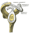
Bankart lesion
Encyclopedia
Injury
-By cause:*Traumatic injury, a body wound or shock produced by sudden physical injury, as from violence or accident*Other injuries from external physical causes, such as radiation injury, burn injury or frostbite*Injury from infection...
of the anterior (inferior
Inferior
Inferior means of lower station, rank, degree, or grade . It may also refer to:* Inferiority complex* An anatomical term of location* Inferior angle of the scapula, in the human skeleton...
) glenoid labrum due to repeated (anterior) shoulder
Shoulder
The human shoulder is made up of three bones: the clavicle , the scapula , and the humerus as well as associated muscles, ligaments and tendons. The articulations between the bones of the shoulder make up the shoulder joints. The major joint of the shoulder is the glenohumeral joint, which...
dislocation. When this happens, a pocket at the front of the glenoid forms that allows the humeral head to dislocate into it. It is an indication for surgery
Surgery
Surgery is an ancient medical specialty that uses operative manual and instrumental techniques on a patient to investigate and/or treat a pathological condition such as disease or injury, or to help improve bodily function or appearance.An act of performing surgery may be called a surgical...
and often accompanied by a Hill-Sachs lesion
Hill-Sachs lesion
A Hill-Sachs lesion, also Hill-Sachs fracture, is a cortical depression in the posterior superior head of the humerus bone. It results from forceful impaction of the humeral head against the anteroinferior glenoid rim when the shoulder is dislocated anteriorly.-Eponym:It is named after Harold...
, damage to the posterior humeral
Humerus
The humerus is a long bone in the arm or forelimb that runs from the shoulder to the elbow....
head.
It is named after Arthur Sydney Blundell Bankart
Arthur Bankart
Arthur Sidney Blundell Bankart was a British orthopaedic surgeon best known for describing the Bankart lesion and Bankart repair for shoulder dislocation.-Biography:...
, an English
English people
The English are a nation and ethnic group native to England, who speak English. The English identity is of early mediaeval origin, when they were known in Old English as the Anglecynn. England is now a country of the United Kingdom, and the majority of English people in England are British Citizens...
orthopaedic surgeon, who lived from 1879-1951.
A bony bankart is a Bankart lesion that includes a fracture
Bone fracture
A bone fracture is a medical condition in which there is a break in the continuity of the bone...
in of the anterior-inferior glenoid cavity
Glenoid cavity
The glenoid cavity is a shallow pyriform, articular surface, which is located on the lateral angle of the scapula. It is directed laterally and forward and articulates with the head of the humerus; it is broader below than above and its vertical diameter is the longest.This cavity forms the...
of the scapula bone.
Imaging
After shoulder dislocations, especially after recurrent anterior dislocations, the osseous structure can be damaged and should be radiologically examined and screened.Normally the inferior portion of the glenoid is wider than the superior portion. This gives the glenoid its pear-shaped appearance. A significant loss of antero-inferior glenoid structure, for example after an anterior dislocation, could lead to a switched appearance. Namely into an inverted pear-shaped glenoid. The rate for recurrent subluxations or dislocations is significantly increased if the glenoid bone appears inverted. Burkhart and DeBeer et al. used this change of morphological appearance as threshold for whether or not a bone grafting procedure (e.g. Latarjet procedure) is required. Additionally to the arthroscopically clearly seen morphological appearance Chuang et al. evaluated the glenoid index via 3D CT scans. The benchmark of the index of 0.75 accurately predicted the requirement of a bone grafting procedure. Therefore the 3D CT scan is a reliable alternative to the invasive arthroscopic examination and can be used as an additional diagnostic tool for preoperative planning and patient counseling.
Conventional radiography has to be applied transaxillary. Without any detached glenoid fragment the axillary view is not able to reliably depict bony lesions in the anterior rim of the glenoid. The Westpoint alignment is a modified transaxillary image, especially qualified for detecting bony Bankart lesions. The patient lies in the ventral position, has the arm abduced 90 degrees, the elbow also 90 degrees flexed and the forearm is hanging over the edge. The cassette is located over the shoulder, the beam direction is cranial medially with the central beam below and medial the acromio-clavicular joint. The position can often be difficult to obtain due to pain after acute dislocation or fear of re-dislocation.
Conventional radiography - X-rays - are a clinically common first choice and well established for depicting bones. But CT has clearly a better resolution and sensitivity concerning detection of bony pathologies. So these two modalities are both potent and clinically reasonable instruments, if used properly. Eiji et al. created stepwise glenoidal defects from 0% up to 46%. The 21% defect, which is reported to cause instability even after surgical Bankart repair, appeared on the Westpoint conventional radiography as a 18.6% defect of the intact glenoid, while on the computed tomography it appeared as a loss of 50% of the width on a single slice across the lower one-fourth of the glenoid.
A tear of the labrum in the sense of a Bankart lesion is an injury of the anterior capsule labrum complex or avulsion fracture of the lower anterior glenoid rim. Involvement of the osseous glenoid makes it per definitionem to a bony Bankart lesion. Through the CT can both the Hill Sachs and the bony Bankart lesion be detected. Even more significance has the arthro-CT, since it can also display the adjacent relevant soft tissue structures, i.e. labrum, joint capsule and gleno-humeral ligaments. An alternative is the MRI. The common planes are axial and coronal, that is perpendicularly to the gleno-humeral joint. Gradient echo sequences and T2 weighted sequences, especially in acute trauma, are preferred. If after the traumatic event it lacks of joint, which serves as a natural contrast agent, the assessment of the labrum and the joint capsule is limited. In these cases an arthrographic MR is advantageous.
Pictures
http://commons.wikimedia.org/wiki/File:BK1.pnghttp://commons.wikimedia.org/wiki/File:BK2.png
http://commons.wikimedia.org/wiki/File:BK3.png
http://commons.wikimedia.org/wiki/File:BK4.png
http://commons.wikimedia.org/wiki/File:BK5.png
External links
- Bankart lesion - orthop.washington.edu
- Bankart lesion - zadeh.co.uk