

Polarized light microscopy
Encyclopedia
Polarized light microscopy can mean any of a number of optical microscopy techniques involving polarized light. Simple techniques include illumination of the sample with polarized light. Directly transmitted light can, optionally, be blocked with a polariser orientated at 90 degrees to the illumination. More complex microscopy techniques which take advantage of polarized light include differential interference contrast microscopy
and interference reflection microscopy
.
These illumination techniques are most commonly used on birefringent samples where the polarized light interacts strongly with the sample and so generating contrast with the background. Polarized light microscopy is used extensively in optical mineralogy
.
Polarized light microscopy is capable of providing information on absorption color and optical path boundaries between minerals of differing refractive indices, in a manner similar to brightfield illumination, but the technique can also distinguish between isotropic and anisotropic substances. Furthermore, the contrast-enhancing technique exploits the optical properties specific to anisotropy and reveals detailed information concerning the structure and composition of materials that are invaluable for identification and diagnostic purposes.
Polarized light is most commonly produced by absorption of light having a set of specific vibration directions in a dichroic medium. Certain natural minerals, such as tourmaline, possess this property, but synthetic films invented by Dr. Edwin H. Land in 1932 soon overtook all other materials as the medium of choice for production of polarized light. Tiny crystallites of iodoquinine sulfate, oriented in the same direction, are embedded in a transparent polymeric film to prevent migration and reorientation of the crystals. Land developed sheets containing polarizing films that were marketed under the trade name of Polaroid®, which has become the accepted generic term for these sheets. Any device capable of selecting polarized light from natural (unpolarized) white light is now referred to as a polar or polarizer, a name first introduced in 1948 by A. F. Hallimond. Today, polarizers are widely used in liquid crystal displays (LCDs), sunglasses, photography, microscopy, and for a myriad of scientific and medical purposes.
There are two polarizing filters in a polarizing microscope - termed the polarizer and analyzer (see Figure 1). The polarizer is positioned beneath the specimen stage usually with its vibration azimuth fixed in the left-to-right, or East-West direction, although most of these elements can be rotated through 360 degrees. The analyzer, usually aligned with a vibration direction oriented North-South, but again rotatable on some microscopes, is placed above the objectives and can be moved in and out of the light path as required. When both the analyzer and polarizer are inserted into the optical path, their vibration azimuths are positioned at right angles to each other. In this configuration, the polarizer and analyzer are said to be crossed, with no light passing through the system and a dark viewfield present in the eyepieces.
For incident light polarized microscopy, the polarizer is positioned in the vertical illuminator and the analyzer is placed above the half mirror. Most rotatable polarizers are graduated to indicate the rotation angle of the transmission azimuth, while analyzers are usually fixed into position (although advanced models can be rotated either 90 or 360 degrees). The polarizer and analyzer are the essential components of the polarizing microscope, but other desirable features include:
Polarized light microscopy can be used both with reflected (incident or epi) and transmitted light. Reflected light is useful for the study of opaque materials such as ceramics, mineral oxides and sulfides, metals, alloys, composites, and silicon wafers (see Figure 3). Reflected light techniques require a dedicated set of objectives that have not been corrected for viewing through the cover glass, and those for polarizing work should also be strain free.
 , where t is the thickness of the sample.
This then leads to a phase difference between the light passing in the two vibration directions of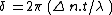 .
.
For example, if the optical path difference is , then the phase difference will be
  , and so the polarisation will be perpendicular to the original, resulting in all of the light passing through the analyser for crossed polars. If the optical path difference is
  , then the phase difference will be
  , and so the polarisation will be parallel to the original. This means that no light will, be able to pass though the analyser which it is now perpendicular to.
The Michel-Levy Chart arises when polarised white light is passed through a birefringent sample. If the sample is of uniform thickness, then only one specific wavelength will meet the above condition described above, and be perpendicular to the direction of the analyser. This means that instead of polychromatic light being viewed at the analyser, one specific wavelength will have been removed. This information can be used in a number of ways:
As the order of the optical path difference increases, then it is more likely that more wavelengths of light will be removed from the spectrum. This results in the appearance of the colour being "washed out", and it becomes more difficult to determine the properties of the sample. This, however, only occurs when the sample is relatively thick when compared to the wavelength of light.
Differential interference contrast microscopy
Differential interference contrast microscopy , also known as Nomarski Interference Contrast or Nomarski microscopy, is an optical microscopy illumination technique used to enhance the contrast in unstained, transparent samples...
and interference reflection microscopy
Interference reflection microscopy
Interference reflection microscopy or IRM is an optical microscopy technique that utilizes polarized light to form an image of an object on a glass surface. The intensity of the signal is a measure of proximity of the object to the glass surface...
.
These illumination techniques are most commonly used on birefringent samples where the polarized light interacts strongly with the sample and so generating contrast with the background. Polarized light microscopy is used extensively in optical mineralogy
Optical mineralogy
Optical mineralogy is the study of minerals and rocks by measuring their optical properties. Most commonly, rock and mineral samples are prepared as thin sections or grain mounts for study in the laboratory with a petrographic microscope...
.
Polarized light microscopy is capable of providing information on absorption color and optical path boundaries between minerals of differing refractive indices, in a manner similar to brightfield illumination, but the technique can also distinguish between isotropic and anisotropic substances. Furthermore, the contrast-enhancing technique exploits the optical properties specific to anisotropy and reveals detailed information concerning the structure and composition of materials that are invaluable for identification and diagnostic purposes.
Basic Properties of Polarized Light
The wave model of light describes light waves vibrating at right angles to the direction of propagation with all vibration directions being equally probable. This is referred to as "common" or "non-polarized" white light. In polarized light there is only one vibration direction (Figure 1). The human eye-brain system has no sensitivity to the vibration directions of light, and polarized light can only be detected by an intensity or color effect, for example, by reduced glare when wearing polarized sun glasses.Polarized light is most commonly produced by absorption of light having a set of specific vibration directions in a dichroic medium. Certain natural minerals, such as tourmaline, possess this property, but synthetic films invented by Dr. Edwin H. Land in 1932 soon overtook all other materials as the medium of choice for production of polarized light. Tiny crystallites of iodoquinine sulfate, oriented in the same direction, are embedded in a transparent polymeric film to prevent migration and reorientation of the crystals. Land developed sheets containing polarizing films that were marketed under the trade name of Polaroid®, which has become the accepted generic term for these sheets. Any device capable of selecting polarized light from natural (unpolarized) white light is now referred to as a polar or polarizer, a name first introduced in 1948 by A. F. Hallimond. Today, polarizers are widely used in liquid crystal displays (LCDs), sunglasses, photography, microscopy, and for a myriad of scientific and medical purposes.
There are two polarizing filters in a polarizing microscope - termed the polarizer and analyzer (see Figure 1). The polarizer is positioned beneath the specimen stage usually with its vibration azimuth fixed in the left-to-right, or East-West direction, although most of these elements can be rotated through 360 degrees. The analyzer, usually aligned with a vibration direction oriented North-South, but again rotatable on some microscopes, is placed above the objectives and can be moved in and out of the light path as required. When both the analyzer and polarizer are inserted into the optical path, their vibration azimuths are positioned at right angles to each other. In this configuration, the polarizer and analyzer are said to be crossed, with no light passing through the system and a dark viewfield present in the eyepieces.
For incident light polarized microscopy, the polarizer is positioned in the vertical illuminator and the analyzer is placed above the half mirror. Most rotatable polarizers are graduated to indicate the rotation angle of the transmission azimuth, while analyzers are usually fixed into position (although advanced models can be rotated either 90 or 360 degrees). The polarizer and analyzer are the essential components of the polarizing microscope, but other desirable features include:
- Specialized Stage - A 360-degree circular rotating specimen stage to facilitate orientation studies with centration of the objectives and stage with the microscope optical axis to make the center of rotation coincide with the center of the field of view. Many stages designed for polarized light microscopy also contain a vernier scale so that rotation angle can be measured to an accuracy of 0.1 degree. For advanced studies of conoscopic images, a universal stage having multiple axes of rotation can also be employed to enable observation of the specimen from any direction.
- Strain Free Objectives - Stress introduced into the glass of an objective during assembly can produce spurious optical effects under polarized light, a factor that could compromise performance. Objectives designed for polarized light observation are distinguished from ordinary objectives with the inscription P, PO, or Pol on the barrel. The performance of an objective is limited by several factors, including the anti-reflection coatings used on lens surfaces, and the refractive properties due to angle of incident light on the front lens. In addition, lens strain can be introduced at the cement junction between elements in a lens group or from a single or group of lenses that has been mounted too tightly in the frame.
- Centerable Revolving Nosepiece - Because the objective optical axis position varies from one assembly to another, many polarized light microscopes are equipped with a specialized nosepiece that contains a centering mechanism for individual objectives. This enables each objective to be centered with respect to the stage and microscope optical axis so that specimen features remain in the center of the viewfield when the stage is rotated through 360 degrees.
- Strain Free Condenser - Condensers designed for polarized light microscopy have several features in common, including the use of strain free lenses. Some condensers are equipped with a receptacle for the polarizer or have the polarizing element mounted directly into the condenser, beneath the aperture diaphragm. Many polarized light condensers have a top lens that can be removed (a swing-lens condenser) from the light path to generate nearly parallel illumination wavefronts for low magnification and birefringence observations.
- Eyepieces - Polarized light microscope eyepieces are fitted with a cross wire reticle (or graticule) to mark the center of the field of view. Often, the cross wire reticle is substituted for a photomicrography reticle that assists in focusing the specimen and composing images with a set of frames bounding the area of the viewfield to be captured either digitally or onto film. Orientation of the eyepiece with respect to the polarizer and analyzer is guaranteed by a point pin that slides into the observation tube sleeve.
- Bertrand Lens - A specialized lens mounted in an intermediate tube or within the observation tubes, a Bertrand lens projects an interference pattern formed at the objective rear focal plane into focus at the microscope image plane. The lens is designed to enable easy examination of the objective rear focal plane, to allow accurate adjustment of the illuminating aperture diaphragm and to view interference figures, similar to the ones presented in Figure 2. Note that in Figure 2(a) and 2(b), the interference patterns represent those observed with a uniaxial crystal in polarized light, while the pattern in Figure 2(c) is typical of a uniaxial crystal with a first order retardation plate inserted into the optical pathway.
- Compensator and Retardation Plates - Many polarized light microscopes contain a slot to allow the insertion of compensators and/or retardation plates between the crossed polarizers, which are used to enhance optical path differences in the specimen. In most modern microscope designs, this slot is placed either in the microscope nosepiece or an intermediate tube positioned between the body and eyepiece tubes. Compensation plates inserted into the slot are then situated between the specimen and the analyzer.
Polarized light microscopy can be used both with reflected (incident or epi) and transmitted light. Reflected light is useful for the study of opaque materials such as ceramics, mineral oxides and sulfides, metals, alloys, composites, and silicon wafers (see Figure 3). Reflected light techniques require a dedicated set of objectives that have not been corrected for viewing through the cover glass, and those for polarizing work should also be strain free.
The Michel-Levy Chart
As polarised light passes through a birefringent sample, the phase difference between the fast and slow directions varies with the thickness, and wavelength of light used. The optical path difference (o.p.d.) is defined as, where t is the thickness of the sample.This then leads to a phase difference between the light passing in the two vibration directions of
.For example, if the optical path difference is
, then the phase difference will be , and so the polarisation will be perpendicular to the original, resulting in all of the light passing through the analyser for crossed polars. If the optical path difference is , then the phase difference will be , and so the polarisation will be parallel to the original. This means that no light will, be able to pass though the analyser which it is now perpendicular to.The Michel-Levy Chart arises when polarised white light is passed through a birefringent sample. If the sample is of uniform thickness, then only one specific wavelength will meet the above condition described above, and be perpendicular to the direction of the analyser. This means that instead of polychromatic light being viewed at the analyser, one specific wavelength will have been removed. This information can be used in a number of ways:
- If the birefringence is known, then the thickness, t, of the sample can be determined
- If the thickness is known, then the birefringence of the sample can be determined
As the order of the optical path difference increases, then it is more likely that more wavelengths of light will be removed from the spectrum. This results in the appearance of the colour being "washed out", and it becomes more difficult to determine the properties of the sample. This, however, only occurs when the sample is relatively thick when compared to the wavelength of light.