

Ossification of maxilla
Encyclopedia
Maxilla
The maxilla is a fusion of two bones along the palatal fissure that form the upper jaw. This is similar to the mandible , which is also a fusion of two halves at the mental symphysis. Sometimes The maxilla (plural: maxillae) is a fusion of two bones along the palatal fissure that form the upper...
is ossified in membrane. Mall 40 and Fawcett 41 maintain that it is ossified from two centers only, one for the maxilla proper and one for the premaxilla.
These centers appear during the sixth week of fetal life and unite in the beginning of the third month, but the suture between the two portions persists on the palate until nearly middle life. Mall states that the frontal process is developed from both centers.
The maxillary sinus appears as a shallow groove on the nasal surface of the bone about the fourth month of fetal life, but does not reach its full size until after the second dentition.
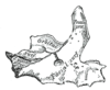
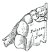
The maxilla was formerly described as ossifying from six centers, viz.,
- one, the orbitonasal, forms that portion of the body of the bone which lies medial to the infraorbital canal, including the medial part of the floor of the orbit and the lateral wall of the nasal cavity;
- a second, the zygomatic, gives origin to the portion which lies lateral to the infraorbital canal, including the zygomatic process;
- from a third, the palatine, is developed the palatine process posterior to the incisive canal together with the adjoining part of the nasal wall;
- a fourth, the premaxillary, forms the incisive bone which carries the incisor teeth and corresponds to the premaxilla of the lower vertebrates;
- a fifth, the nasal, gives rise to the frontal process and the portion above the canine tooth;
- and a sixth, the infravomerine, lies between the palatine and premaxillary centers and beneath the vomer; this center, together with the corresponding center of the opposite bone, separates the incisive canals from each other.
Changes produced in the maxilla by age
At birth the transverse and antero-posterior diameters of the bone are each greater than the vertical.The frontal process is well-marked and the body of the bone consists of little more than the alveolar process, the teeth sockets reaching almost to the floor of the orbit.
The maxillary sinus presents the appearance of a furrow on the lateral wall of the nose. In the adult the vertical diameter is the greatest, owing to the development of the alveolar process and the increase in size of the sinus.
In old age the bone reverts in some measure to the infantile condition; its height is diminished, and after the loss of the teeth the alveolar process is absorbed, and the lower part of the bone contracted and reduced in thickness.
See also
- Changes produced in the mandible by ageChanges produced in the mandible by agethumb|Fig. 1: At birth.thumb|Fig. 2: In childhood.thumb|Fig. 3: In the adult.thumb|Fig. 4: In old age. Side view of the mandible at different periods of life....