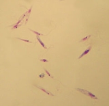
Leishmania tropica
Encyclopedia
Leishmania tropica is a species
of flagellate
parasites
that infects humans and rodent
s. It can cause a disease
called oriental sore which is a form of cutaneous leishmaniasis
.Leishmania tropica is a single-celled trypanosome parasite responsible for causing cutaneous Leishmaniasis.[1] Leishmaniasis is found in approximately 88 countries world-wide.¬[1][3] Its prevalence in several countries around the world is apparent by the many names it has adopted: "Baghdad Boil", "Jericho buttons", and "Oriental sore", to name a few.[1][2] Cutaneous Leishmaniasis develops at the site of a sand fly bite. The sand fly, which carries the promastigote stage of L. tropica in its blood, is the vector.
Diagnostic findings
Microscopy
Isolation of the organism in culture (using for example the diphasic NNN medium) or in experimental animals (hamsters) constitutes another method of parasitilogic confirmation of the diagnosis, and in addition can provide material for further investigations (e.g., isoenzyme analysis).[8] Antibody detection can prove useful in visceral leishmaniasis but is of limited value in cutaneous disease, where most patients do not develop a significant antibody response. In addition, cross reactivity can occur with Trypanosoma cruzi, a fact to consider when investigating Leishmania antibody response in patients who have been in Central or South America.[8] Other diagnostic techniques exist that allow parasite detection and/or species identification using biochemical (isoenzymes), immunologic (immunoassays), and molecular (PCR) approaches. Such techniques, however, are not readily available in general diagnostic laboratories.[8]
2. Immunity as a result of the natural course of the disease is 97% to 98% effective. Some native peoples deliberately inoculate their children on a part of their bodies that are normally covered by clothing. This practice prevented a child from later developing a disfiguring scar on an exposed part of their body.[6]
.
2. United States of America. Department of Health and Human Services. Center for Disease Control and Prevention. Leishmaniasis – General Information. July 2009. 15 Nov. 2010.
3. United States of America. Department of Health and Human Services. Center for Disease Control and Prevention. Leishmaniasis - Biology. July 2009. 15 Nov. 2010.
4. James, William D.; Berger, Timothy G.; et al. (2006). Andrews' Diseases of the Skin: clinical Dermatology. Saunders Elsevier
5. Sehgal, Ravinder. “Blood and Tissue Flagellates” San Francisco State University, San Francisco, CA. 14 September. 2010.
6. Schmidt G.D. and Roberts L.S. 2009. Foundations of Parasitology. 8th ed. McGraw-Hill, Dubuque, IA, p. 76-81.
7. Perlin, David and Cohen, Ann (2002). The Complete Idiot's Guide to Dangerous Diseases and Epidemics. 1st ed. Indianapolis, IN: Marie Butler-Knight, 2002.
8. United States of America. Department of Health and Human Services. Center for Disease Control and Prevention. Leishmaniasis. July 2009. 15 Nov. 2010.
Species
In biology, a species is one of the basic units of biological classification and a taxonomic rank. A species is often defined as a group of organisms capable of interbreeding and producing fertile offspring. While in many cases this definition is adequate, more precise or differing measures are...
of flagellate
Flagellate
Flagellates are organisms with one or more whip-like organelles called flagella. Some cells in animals may be flagellate, for instance the spermatozoa of most phyla. Flowering plants do not produce flagellate cells, but ferns, mosses, green algae, some gymnosperms and other closely related plants...
parasites
Parasitism
Parasitism is a type of symbiotic relationship between organisms of different species where one organism, the parasite, benefits at the expense of the other, the host. Traditionally parasite referred to organisms with lifestages that needed more than one host . These are now called macroparasites...
that infects humans and rodent
Rodent
Rodentia is an order of mammals also known as rodents, characterised by two continuously growing incisors in the upper and lower jaws which must be kept short by gnawing....
s. It can cause a disease
Disease
A disease is an abnormal condition affecting the body of an organism. It is often construed to be a medical condition associated with specific symptoms and signs. It may be caused by external factors, such as infectious disease, or it may be caused by internal dysfunctions, such as autoimmune...
called oriental sore which is a form of cutaneous leishmaniasis
Cutaneous leishmaniasis
Cutaneous leishmaniasis is the most common form of leishmaniasis. It is a skin infection caused by a single-celled parasite that is transmitted by sandfly bites...
.Leishmania tropica is a single-celled trypanosome parasite responsible for causing cutaneous Leishmaniasis.[1] Leishmaniasis is found in approximately 88 countries world-wide.¬[1][3] Its prevalence in several countries around the world is apparent by the many names it has adopted: "Baghdad Boil", "Jericho buttons", and "Oriental sore", to name a few.[1][2] Cutaneous Leishmaniasis develops at the site of a sand fly bite. The sand fly, which carries the promastigote stage of L. tropica in its blood, is the vector.
Life cycle
Leishmaniasis is transmitted by the bite of infected female phlebotomine sandflies. The life cycle of Leishmania Tropica is identical to that of other related parasites of the same genus and includes both an amastigote and a promastigote stage. The sand flies inject the infective stage of promastigote.[1] The promastigote stage is considered to be part of the infective stage, in which a sand fly infects a host with the parasite through feeding.[1] The amastigote is part of the tissue stage in which the parasite transforms after being engulfed by a macrophage.[1]Pathogenesis
The sandflies inject the infective stage of promastigote from their proboscis (feeding structure) during blood meals.[4] At this point, they are in the promastigote stage, which is from the time that they are ingested by a sand fly until they are phagocytized by a macrophage. Promastigotes that reach the puncture wound are phagocytized by macrophages and other mononuclear phagocytic cells.[4] Progmastigotes then transform inside of infected cells into the tissue stage of the parasite, the amastigote. The amastigote stage multiplies by simple division and proceeds to infect other mononuclear phagocytic cells.[4] In a human host, this parasite is responsible for transmitting and infecting its victims with cutaneous leishmaniasis. Sandflies become infected by ingesting infected cells during blood meals.[4] In sandflies, amastigotes transform into promastigotes, develop in the gut and migrate to the proboscis.[4]Disease
There are several forms of Leishmaniasis which are caused by various species of Leishmania. L. tropica is solely responsible for causing cutaneous disease, including the characteristic sore at the site of a sand fly bite. The sore is a result of the extreme immune inflammatory response initiated by a host after being exposed to the parasite. Other Leishmania species are responsible for visceral and mucocutaneous leishmaniasis.Microscopic appearance
L. Tropica has both the promastigote and the amastigote stage. The promastigote is a motile flagellate that is about 10-20 micrometers long.[6] The amastigotes are similar to those of other leshmanias. Amastigotes are spheroid to ovoid in shape and are 2.5- 5.0 micrometers wide but some can be smaller. It contains a nucleus and a kinetoplast and the cytoplasm is vacuolated.Origins and evolution
Cutaneous Leishmania that is caused by L. tropica is found in the old world: Mediterranean, Middle East, Africa, and India.[6] It is believed that the transport of slaves to the western world from Africa through the Middle East and Asia spread Leishmania species into previously uncontaminated areas.[6] L. tropica is most similar to L. major due to its life cycle but are found in different locations and have different reservoir and intermediate host.Prevention
The best method is insecticide spraying to kill the vector, or transport host the san fly.[5] Travelers should wear protective clothing and use insect repellent. Bed nets and screen doors and windows should be used as well. The netting must be very fine in order to be effective, as sand flies are about one third the size of mosquitoes.[7]Diagnosis
Laboratory Diagnosis-Examination of Giemsa stained slides of the relevant tissue is still the technique most commonly used to detect the parasite.Diagnostic findings
Microscopy
Isolation of the organism in culture (using for example the diphasic NNN medium) or in experimental animals (hamsters) constitutes another method of parasitilogic confirmation of the diagnosis, and in addition can provide material for further investigations (e.g., isoenzyme analysis).[8] Antibody detection can prove useful in visceral leishmaniasis but is of limited value in cutaneous disease, where most patients do not develop a significant antibody response. In addition, cross reactivity can occur with Trypanosoma cruzi, a fact to consider when investigating Leishmania antibody response in patients who have been in Central or South America.[8] Other diagnostic techniques exist that allow parasite detection and/or species identification using biochemical (isoenzymes), immunologic (immunoassays), and molecular (PCR) approaches. Such techniques, however, are not readily available in general diagnostic laboratories.[8]
Treatment
1. Drugs: pentavalent antimony, amphotericin B, miltefosine can be used. These are toxic and only warranted for L. braziliensis and L. donovani or severe cutaneous cases. There are possible complications such as post kala azar dermal lesions.[5]2. Immunity as a result of the natural course of the disease is 97% to 98% effective. Some native peoples deliberately inoculate their children on a part of their bodies that are normally covered by clothing. This practice prevented a child from later developing a disfiguring scar on an exposed part of their body.[6]
External links
1. Dictionary - Definition of Leishmaniasis. 15 Nov. 2010. 15 Nov. 20102. United States of America. Department of Health and Human Services. Center for Disease Control and Prevention. Leishmaniasis – General Information. July 2009. 15 Nov. 2010
3. United States of America. Department of Health and Human Services. Center for Disease Control and Prevention. Leishmaniasis - Biology. July 2009. 15 Nov. 2010
4. James, William D.; Berger, Timothy G.; et al. (2006). Andrews' Diseases of the Skin: clinical Dermatology. Saunders Elsevier
5. Sehgal, Ravinder. “Blood and Tissue Flagellates” San Francisco State University, San Francisco, CA. 14 September. 2010.
6. Schmidt G.D. and Roberts L.S. 2009. Foundations of Parasitology. 8th ed. McGraw-Hill, Dubuque, IA, p. 76-81.
7. Perlin, David and Cohen, Ann (2002). The Complete Idiot's Guide to Dangerous Diseases and Epidemics. 1st ed. Indianapolis, IN: Marie Butler-Knight, 2002.
8. United States of America. Department of Health and Human Services. Center for Disease Control and Prevention. Leishmaniasis. July 2009. 15 Nov. 2010

