

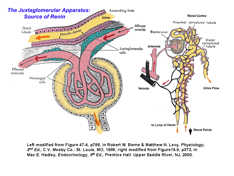
Juxtaglomerular apparatus
Encyclopedia
Kidney
The kidneys, organs with several functions, serve essential regulatory roles in most animals, including vertebrates and some invertebrates. They are essential in the urinary system and also serve homeostatic functions such as the regulation of electrolytes, maintenance of acid–base balance, and...
, which regulates the function of each nephron
Nephron
The renal tubule is the portion of the nephron containing the tubular fluid filtered through the glomerulus. After passing through the renal tubule, the filtrate continues to the collecting duct system, which is not part of the nephron....
. The juxtaglomerular apparatus is named for its proximity to the glomerulus
Glomerulus
A glomerulus is a capillary tuft that is involved in the first step of filtering blood to form urine.A glomerulus is surrounded by Bowman's capsule, the beginning component of nephrons in the vertebrate kidney. A glomerulus receives its blood supply from an afferent arteriole of the renal...
: it is found between the vascular pole
Vascular pole
The vascular pole is a location of the glomerulus. At the vascular pole, the afferent arterioles and efferent arterioles enter the Bowman's capsule.The urinary pole is at the other end....
of the renal corpuscle
Renal corpuscle
In the kidney, a renal corpuscle is the initial blood-filtering component of a nephron. It consists of two structures: a glomerulus and a Bowman's capsule. The glomerulus is a small tuft of capillaries containing two cell types. Endothelial cells, which have large fenestrae, are not covered by...
and the returning distal convoluted tubule
Distal convoluted tubule
The distal convoluted tubule is a portion of kidney nephron between the loop of Henle and the collecting duct system.- Physiology :It is partly responsible for the regulation of potassium, sodium, calcium, and pH...
of the same nephron. This location is critical to its function in regulating renal blood flow and glomerular filtration rate. The three cellular components of the apparatus are the macula densa
Macula densa
In the kidney, the macula densa is an area of closely packed specialized cells lining the wall of the distal tubule at the point of return of the nephron to the vascular pole of its parent glomerulus, ....
, extraglomerular mesangial cells, and juxtaglomerular cell
Juxtaglomerular cell
The juxtaglomerular cells are cells in the kidney that synthesize, store, and secrete the enzyme renin. They are specialized smooth muscle cells in the wall of the afferent arteriole that delivers blood to the glomerulus...
s (juxtaglomerular cells are not granular cells but are granulated as they release Renin).
Cells of the Juxtaglomerular Apparatus
There are 3 different types of cells in the Juxtaglomerular Apparatus: Granular Cells, Macula Densa Cells, and Mesangial Cells.Granular Cells
Granular cells are modified pericytes of glomerular arterioles. They are also known as Juxtaglomerular cells..The Juxtaglomerular cells secrete renin
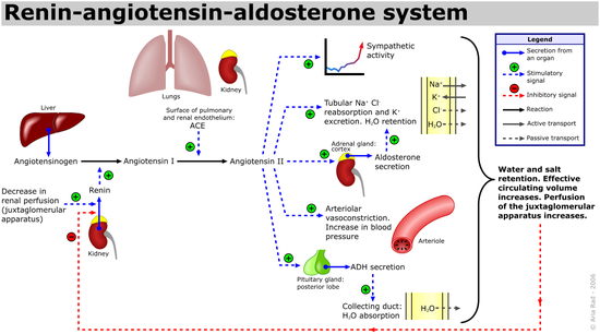
Renin
Renin , also known as an angiotensinogenase, is an enzyme that participates in the body's renin-angiotensin system -- also known as the Renin-Angiotensin-Aldosterone Axis -- that mediates extracellular volume , and arterial vasoconstriction...
in response to:
- Beta1 adrenergic stimulation
- Decrease in renal perfusion pressure (detected directly by the granular cells)
- Decrease in NaCl absorption in the Macula Densa (often due to a decrease in glomerular filtration rate, or GFR, causing slower filtrate movement through the proximal tubule and thus more time for reabsorption. This results in a lower NaCl concentration by the time the filtrate reaches the Macula Densa).
Macula Densa Cells
Macula densa cells are columnar epithelium thickening of the distal tubule. The macula densa senses sodium chlorideSodium chloride
Sodium chloride, also known as salt, common salt, table salt or halite, is an inorganic compound with the formula NaCl. Sodium chloride is the salt most responsible for the salinity of the ocean and of the extracellular fluid of many multicellular organisms...
concentration in the distal tubule of the kidney and secretes a locally active (paracrine) vasopressor which acts on the adjacent afferent arteriole to decrease glomerular filtration rate (GFR), as part of the tubuloglomerular feedback loop. Specifically, excessive filtration at the glomerulus or inadequate sodium uptake in the proximal tubule / thick ascending loop of Henle brings fluid to the distal convoluted tubule that has an abnormally high concentration of sodium. Na/Cl cotransporters move sodium into the cells of the macula densa. The macula densa cells do not have enough basolateral Na/K ATPases to excrete this added sodium, so the cell's osmolarity increases. Water flows into the cell to bring the osmolarity back down, causing the cell to swell. When the cell swells, a stretch-activated non-selective anion channel is opened on the basolateral surface. ATP escapes through this channel and is subsequently converted to adenosine. Adenosine vasoconstricts the afferent arteriole via A1 receptors and vasodilates (to a lesser degree) efferent arterioles via A2 receptors which decreases GFR. Also, adenosine inhibits renin release in JG cells via A2 receptors on JG cells using Gi pathway. Also, when macula densa cells detect higher concentrations of Na and Cl they inhibit Nitric Oxide Synthetase (decreasing renin release) with an unknown pathway.
A decrease in GFR means less solute in the tubular lumen. As the filtrate reaches the macula densa, less NaCl is re-absorbed. The macula densa cells detect lower concentrations in Na and Cl and upregulate Nitric Oxide Synthetase (NOS). NOS creates NO which catalyses the formation of prostaglandins. These prostaglandins diffuse to the granular cells and activate a prostaglandin specific Gs receptor. This receptor activates adenylate cyclase which increases levels of cAMP. cAMP augments renin release. Prostaglandins and NO also makes a vasadilator effect on afferent arteriol but this doesn't happen on efferent arteriol due to renin release.
Mesangial cells
Mesangial cells are structural cells in the glomerulus that under normal conditions serve as anchors for the glomerular capillaries. The mesangial cells within the glomerulus communicate with mesangial cells outside the glomerulus (extraglomerular mesangial cells), and it is the latter cells that form part of the juxtaglomerular apparatus. These cells form a syncytiumSyncytium
In biology, a syncytium is a large cell-like structure; filled with cytoplasm and containing many nuclei. Most cells in eukaryotic organisms have a single nucleus; syncytia are specialized forms used by various organisms.The term may also refer to cells that are connected by specialized membrane...
and are connected with glomerular mesangial cells via gap junctions.
The function of the extraglomerular mesangial cells remains somewhat mysterious. They contain actin
Actin
Actin is a globular, roughly 42-kDa moonlighting protein found in all eukaryotic cells where it may be present at concentrations of over 100 μM. It is also one of the most highly-conserved proteins, differing by no more than 20% in species as diverse as algae and humans...
and myosin
Myosin
Myosins comprise a family of ATP-dependent motor proteins and are best known for their role in muscle contraction and their involvement in a wide range of other eukaryotic motility processes. They are responsible for actin-based motility. The term was originally used to describe a group of similar...
, allowing them to contract when stimulated by renal sympathetic nerves
Sympathetic nervous system
The sympathetic nervous system is one of the three parts of the autonomic nervous system, along with the enteric and parasympathetic systems. Its general action is to mobilize the body's nervous system fight-or-flight response...
, which may provide a way for the sympathetic nervous system to modulate the actions of the juxtaglomerular apparatus. The latest thinking is that in times of great sympathetic discharge [i.e. during periods when the blood pressure is low, e.g. from blood loss ], mesangial contraction reduces the surface area of the glomerulae, thus reducing glomerular filtration and saving excess fluid from being lost into the urine. In addition, extraglomerular mesangial cells are strategically positioned between the macula densa and the afferent arteriole, and may mediate signalling between these two structures.