
Indocyanine green
Encyclopedia
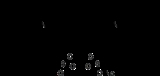
Indocyanine green is a cyanine
dye
used in medical diagnostics. It is used for determining cardiac output, hepatic function, and liver blood flow, and for ophthalmic angiography. It has a peak spectral absorption at about 800 nm. These infrared
frequencies penetrate retinal layers, allowing ICG angiography to image deeper patterns of circulation than Fluorescein angiography
. ICG binds tightly to plasma proteins and becomes confined to the vascular system. ICG has a half-life
of 150 to 180 seconds and is removed from circulation exclusively by the liver to bile juice.
Indocyanine green (ICG) is a fluorescent dye which is used in medicine as an indicator substance (e.g. for photometric hepatic function diagnostics and fluorescence angiography) in cardiac, circulatory, hepatic and ophthalmic conditions.(Definition of indocyanine green, National Cancer Institute). It is administered intravenously and, depending on liver performance, is eliminated from the body with a half life of approx. 3-4 minutes. ICG sodium salt is normally available in powder form and can be dissolved in various solvents; 5% (<5% depending on batch) sodium iodide is usually added to ensure better solubility. The sterile lyophilisate of a water-ICG solution is approved many European countries and the United States under the name ICG-Pulsion® (manufacturer: Pulsion) and IC-Green® (manufacturer: Akorn) as a diagnostic for intravenous use.
History
ICG was developed in the Second World War as a colouring agent in photography and tested in 1957 at the Mayo Clinic for use in human medicine. After being granted FDA approval in 1959, ICG was initially used primarily in hepatic function diagnostics and later in cardiology. In 1964, S. Schilling was able to determine renal blood flow using indocyanine green. From 1969, ICG was also used in the research and diagnosis of subretinal processes in the eye (in the choroid). In the years since 1980, the development of new types of cameras and better film material or new photometric measuring devices has cleared away many technical difficulties. In the mean time, the use of ICG in medicine (and especially in fluorescent angiography in ophthalmology) has become established as standard. A distinction is therefore also made, when describing fluorescent angiography, between NA fluorescent angiography and ICGA / ICG fluorescent angiography. Around 3,000 scientific papers on ICG have now been published worldwide.
Optical properties
The absorption and fluorescence spectrum of ICG is in the near infrared region. Both depend largely on the solvent used and the concentration. ICG absorbs mainly between 600 nm and 900 nm and emits fluorescence between 750 nm and 950 nm. The large overlapping of the absorption and fluorescence spectra leads to a marked reabsorption of the fluorescence by ICG itself. The fluorescence spectrum is very wide. Its maximum values are approx. 810 nm in water and approx. 830 nm in blood. For medical applications based on absorption, the maximum absorption at approx. 800 nm (in blood plasma at low concentrations) is important. In combination with fluorescence detection, lasers with a wavelength of around 780 nm are used. At this wavelength, ICG still absorbs very well and yet it is still technically possible to suppress the excitation light in order to detect the fluorescence.
Toxicity and side-effects
Indocyanine green is metabolised microsomally in the liver and only excreted via the liver and bile ducts; since it is not absorbed by the intestinal mucous membrane, the toxicity can be classified as low. Administration is not without risks during pregnancy. It has been known since September 2007 that ICG decomposes into toxic waste materials under the influence of UV light, creating a number of still unknown substances. A study published in February 2008, however, shows that ICG (the substance without UV effect) is basically, as such, of only minor toxicity. The intravenous LD50 values measured in animals are 60 mg/kg in mice and 87 mg/kg in rats. Occasionally – in one out of 42,000 cases – slight side-effects occur in humans such as sore throats and hot flushes. Effects such as anaphylactic shock, hypotension, tachycardia, dyspnoea and urticaria only occurred in individual cases; the risk of severe side-effects rises in patients with chronic kidney impairment. The frequencies of mild, moderate and severe side-effects were only 0.15%, 0.2% and 0.05%; the rate of deaths is 1:333,333. For the competitor substance fluorescein, the proportion of people with side-effects is 4.8% and the death rate is 1:222,222.
ICG angiography in ophthalmology.
Because the preparation contains sodium iodide, a test must be carried out for iodine intolerance. Because around 5% of iodide is added, the iodine content of a 25 mg ampoule is 0.93 mg. In comparison, preparations for a bone marrow CT (140 ml) contain 300 mg/ml and for a corona angiography (200 ml) 350 mg/ml of iodine.
Indocyanine green or ICG has the ability to bind 98% to plasma proteins – 80% to globulins and 20% to alpha-lipoprotein and albumin – and thus, in comparison with fluorescein as a marker, has a lower leakage (slower emergence of dye from the vessels, extravasally).^ Ophthalmic Diagnostic Photography; Indocyanine Green (ICG) Angiography University of Iowa Health Care). Because of the plasma protein binding, ICG stays for up to 20–30 minutes in the vessels (intravasally). When the eye is examined, it thus stays for a long time in tissues with a higher blood flow, such as the choroid and the blood vessels of the retina.In ophthalmology, the ICG angiography is used for an angiography of the ocular fundus in the following cases:
• If there is a suspicion of retinal damage with poorly defined edges and if there is bleeding
• In basic diagnostics, if there is a suspicion of a particular form of AMD
• If there is a suspicion of choroidal neovascularisation (CNV)
• If there is a suspicion of polypoidal choroidal vasculopathy (PCV)
• If there is a suspicion of retinal angiomatous proliferation (RAP)
• If there is a suspicion of choroidal melanoma
• If there is a suspicion of choroidal haemangioma
• If there is a suspicion of choroidal metastases
• In individual cases for extended diagnostics or documentation (differential diagnostics)
ICG diagnostics for the non-invasive monitoring of liver or splanchnic perfusion
Because of the high binding rate of indocyanine green to plasma proteins, the substance allows liver / splanchnic perfusion to be measured by monitoring the changes in the ICG-PDR (ICG plasma disappearance rate). This method is therefore suitable as a parameter for predicting the probability of survival of intensive-care surgical patients.Since reduced PDR values can be measured in about two thirds of intensive-care surgical patients in whom expanded haemodynamic monitoring is indicated, it is clear that there is a significantly increased mortality rate here. Regular monitoring of the ICG-PDR (generally twice a day) helps to reveal restrictions in liver / splanchnic perfusion at an early stage. After intravenous ICG administration, the plasma disappearance rate is measured using an external monitoring device.
Perfusion diagnostics of tissues and organs using ICG
ICG is used as a marker in the assessment of the perfusion of tissues and organs in many areas of medicine. The light needed for the excitation of the fluorescence is generated by a near infrared light source which is attached directly to a camera. A digital video camera allows the absorption of the ICG fluorescence to be recorded in real time, which means that perfusion can be assessed and documented. This is used in the following cases: • Plastic surgery: skin and muscle transplants; determination of amputation level • Abdominal surgery: gastrointestinal anastomosis • General surgery: wound healing and ulcers • Internal medicine: diabetic extremities • Heart surgery: aortocoronary bypasses In addition, ICG can also be used as a tracer in cerebral perfusion diagnostics. In the case of stroke patients, monitoring in the recovery phase seems to be achievable by measurement of both the ICG absorption and the fluorescence in everyday clinical conditions.
ICG-supported navigation for sentinel lymph node biopsy with tumours
Sentinel lymph node biopsy (SLB or SLN biopsy) allows selective, minimally invasive access for assessment of the regional lymph node status with malignant tumours. The first draining lymph note, the "sentinel", represents an existing or non-existing tumour of an entire lymph node region. The method has been validated using radionuclides and/or blue dye for breast cancer, malignant melanoma and also gastrointestinal tumours and gives a good detection rate and sensitivity. For the SLB, a reduced mortality has been observed in comparison with complete lymph node dissection, but the methods have disadvantages with regard to availability, application and disposal of the radionuclide and the risk of anaphylaxis (up to 1%) for the blue dye. Indocyanine green (ICG), because of its NIR (near-infrared) fluorescence and previous toxicity investigations, was evaluated in this investigation as a new, alternative method for SLB with regard to the clinical application of the transcutaneous navigation and lymph vessel visualisation and SLN detection. ICG fluorescence navigation achieves high rates of detection and sensitivity in comparison with the conventional methods. Taking into account the learning curve required, the new, alternative method offers a combination of lymphography and SLB and the possibility of carrying out an SLB without the need for radioactive substances for solitary tumours
Diagnosis of rheumatic diseases
The Physikalisch-Technische Bundesanstalt and mivenion GmbH developed an imaging process, known as "Rheumascan", for the diagnosis of microcirculatory problems in the hands. This imaging process can be used with arthritic and rheumatoid diseases. ICG is used as the marker for the microcirculatory disorder. Rheumascan was presented for the first time in 2009 at the European Congress of Radiology.
Ongoing research
Research in the area of ICG has by no means been completed at the current time. In particular, it seems that, under certain circumstances, such as the right laser strength and particular wavelengths, ICG may have the capacity to trigger a photodynamic reaction. ICG could thus also be used in the medium term as a PDT therapy. On the other hand, ICG can be used very universally in the area of (perfusion) diagnostics, and further research is therefore also being carried out here.
(the world's largest manufacturer of ICG)
Indocyanine green angiography in chorioretinal diseases: indications and interpretation: an evidence-based update. In: Ophthalmology. Vol. 110, p. 15–21. PMID 12511340
]ICG website of Akorn
Verband der Deutschen Ophthalmologen (Association of German Ophthalmologists)
http://omlc.ogi.edu/spectra/icg/Optical Absorption of Indocyanine Green (ICG )
Cyanine
Cyanine is a non-systematic name of a synthetic dye family belonging to polymethine group. Cyanines have many uses as fluorescent dyes, particularly in biomedical imaging...
dye
Dye
A dye is a colored substance that has an affinity to the substrate to which it is being applied. The dye is generally applied in an aqueous solution, and requires a mordant to improve the fastness of the dye on the fiber....
used in medical diagnostics. It is used for determining cardiac output, hepatic function, and liver blood flow, and for ophthalmic angiography. It has a peak spectral absorption at about 800 nm. These infrared
Infrared
Infrared light is electromagnetic radiation with a wavelength longer than that of visible light, measured from the nominal edge of visible red light at 0.74 micrometres , and extending conventionally to 300 µm...
frequencies penetrate retinal layers, allowing ICG angiography to image deeper patterns of circulation than Fluorescein angiography
Fluorescein angiography
Intravenous Fluorescein angiography or fluorescent angiography is a technique for examining the circulation of the retina using the dye tracing method...
. ICG binds tightly to plasma proteins and becomes confined to the vascular system. ICG has a half-life
Half-life
Half-life, abbreviated t½, is the period of time it takes for the amount of a substance undergoing decay to decrease by half. The name was originally used to describe a characteristic of unstable atoms , but it may apply to any quantity which follows a set-rate decay.The original term, dating to...
of 150 to 180 seconds and is removed from circulation exclusively by the liver to bile juice.
Indocyanine green (ICG) is a fluorescent dye which is used in medicine as an indicator substance (e.g. for photometric hepatic function diagnostics and fluorescence angiography) in cardiac, circulatory, hepatic and ophthalmic conditions.(Definition of indocyanine green, National Cancer Institute). It is administered intravenously and, depending on liver performance, is eliminated from the body with a half life of approx. 3-4 minutes. ICG sodium salt is normally available in powder form and can be dissolved in various solvents; 5% (<5% depending on batch) sodium iodide is usually added to ensure better solubility. The sterile lyophilisate of a water-ICG solution is approved many European countries and the United States under the name ICG-Pulsion® (manufacturer: Pulsion) and IC-Green® (manufacturer: Akorn) as a diagnostic for intravenous use.
History
ICG was developed in the Second World War as a colouring agent in photography and tested in 1957 at the Mayo Clinic for use in human medicine. After being granted FDA approval in 1959, ICG was initially used primarily in hepatic function diagnostics and later in cardiology. In 1964, S. Schilling was able to determine renal blood flow using indocyanine green. From 1969, ICG was also used in the research and diagnosis of subretinal processes in the eye (in the choroid). In the years since 1980, the development of new types of cameras and better film material or new photometric measuring devices has cleared away many technical difficulties. In the mean time, the use of ICG in medicine (and especially in fluorescent angiography in ophthalmology) has become established as standard. A distinction is therefore also made, when describing fluorescent angiography, between NA fluorescent angiography and ICGA / ICG fluorescent angiography. Around 3,000 scientific papers on ICG have now been published worldwide. Optical properties
The absorption and fluorescence spectrum of ICG is in the near infrared region. Both depend largely on the solvent used and the concentration. ICG absorbs mainly between 600 nm and 900 nm and emits fluorescence between 750 nm and 950 nm. The large overlapping of the absorption and fluorescence spectra leads to a marked reabsorption of the fluorescence by ICG itself. The fluorescence spectrum is very wide. Its maximum values are approx. 810 nm in water and approx. 830 nm in blood. For medical applications based on absorption, the maximum absorption at approx. 800 nm (in blood plasma at low concentrations) is important. In combination with fluorescence detection, lasers with a wavelength of around 780 nm are used. At this wavelength, ICG still absorbs very well and yet it is still technically possible to suppress the excitation light in order to detect the fluorescence. Toxicity and side-effects
Indocyanine green is metabolised microsomally in the liver and only excreted via the liver and bile ducts; since it is not absorbed by the intestinal mucous membrane, the toxicity can be classified as low. Administration is not without risks during pregnancy. It has been known since September 2007 that ICG decomposes into toxic waste materials under the influence of UV light, creating a number of still unknown substances. A study published in February 2008, however, shows that ICG (the substance without UV effect) is basically, as such, of only minor toxicity. The intravenous LD50 values measured in animals are 60 mg/kg in mice and 87 mg/kg in rats. Occasionally – in one out of 42,000 cases – slight side-effects occur in humans such as sore throats and hot flushes. Effects such as anaphylactic shock, hypotension, tachycardia, dyspnoea and urticaria only occurred in individual cases; the risk of severe side-effects rises in patients with chronic kidney impairment. The frequencies of mild, moderate and severe side-effects were only 0.15%, 0.2% and 0.05%; the rate of deaths is 1:333,333. For the competitor substance fluorescein, the proportion of people with side-effects is 4.8% and the death rate is 1:222,222. ICG angiography in ophthalmology.
Because the preparation contains sodium iodide, a test must be carried out for iodine intolerance. Because around 5% of iodide is added, the iodine content of a 25 mg ampoule is 0.93 mg. In comparison, preparations for a bone marrow CT (140 ml) contain 300 mg/ml and for a corona angiography (200 ml) 350 mg/ml of iodine. Indocyanine green or ICG has the ability to bind 98% to plasma proteins – 80% to globulins and 20% to alpha-lipoprotein and albumin – and thus, in comparison with fluorescein as a marker, has a lower leakage (slower emergence of dye from the vessels, extravasally).^ Ophthalmic Diagnostic Photography; Indocyanine Green (ICG) Angiography University of Iowa Health Care). Because of the plasma protein binding, ICG stays for up to 20–30 minutes in the vessels (intravasally). When the eye is examined, it thus stays for a long time in tissues with a higher blood flow, such as the choroid and the blood vessels of the retina.In ophthalmology, the ICG angiography is used for an angiography of the ocular fundus in the following cases:
• If there is a suspicion of retinal damage with poorly defined edges and if there is bleeding
• In basic diagnostics, if there is a suspicion of a particular form of AMD
• If there is a suspicion of choroidal neovascularisation (CNV)
• If there is a suspicion of polypoidal choroidal vasculopathy (PCV)
• If there is a suspicion of retinal angiomatous proliferation (RAP)
• If there is a suspicion of choroidal melanoma
• If there is a suspicion of choroidal haemangioma
• If there is a suspicion of choroidal metastases
• In individual cases for extended diagnostics or documentation (differential diagnostics)
ICG diagnostics for the non-invasive monitoring of liver or splanchnic perfusion
Because of the high binding rate of indocyanine green to plasma proteins, the substance allows liver / splanchnic perfusion to be measured by monitoring the changes in the ICG-PDR (ICG plasma disappearance rate). This method is therefore suitable as a parameter for predicting the probability of survival of intensive-care surgical patients.Since reduced PDR values can be measured in about two thirds of intensive-care surgical patients in whom expanded haemodynamic monitoring is indicated, it is clear that there is a significantly increased mortality rate here. Regular monitoring of the ICG-PDR (generally twice a day) helps to reveal restrictions in liver / splanchnic perfusion at an early stage. After intravenous ICG administration, the plasma disappearance rate is measured using an external monitoring device. Perfusion diagnostics of tissues and organs using ICG
ICG is used as a marker in the assessment of the perfusion of tissues and organs in many areas of medicine. The light needed for the excitation of the fluorescence is generated by a near infrared light source which is attached directly to a camera. A digital video camera allows the absorption of the ICG fluorescence to be recorded in real time, which means that perfusion can be assessed and documented. This is used in the following cases: • Plastic surgery: skin and muscle transplants; determination of amputation level • Abdominal surgery: gastrointestinal anastomosis • General surgery: wound healing and ulcers • Internal medicine: diabetic extremities • Heart surgery: aortocoronary bypasses In addition, ICG can also be used as a tracer in cerebral perfusion diagnostics. In the case of stroke patients, monitoring in the recovery phase seems to be achievable by measurement of both the ICG absorption and the fluorescence in everyday clinical conditions. ICG-supported navigation for sentinel lymph node biopsy with tumours
Sentinel lymph node biopsy (SLB or SLN biopsy) allows selective, minimally invasive access for assessment of the regional lymph node status with malignant tumours. The first draining lymph note, the "sentinel", represents an existing or non-existing tumour of an entire lymph node region. The method has been validated using radionuclides and/or blue dye for breast cancer, malignant melanoma and also gastrointestinal tumours and gives a good detection rate and sensitivity. For the SLB, a reduced mortality has been observed in comparison with complete lymph node dissection, but the methods have disadvantages with regard to availability, application and disposal of the radionuclide and the risk of anaphylaxis (up to 1%) for the blue dye. Indocyanine green (ICG), because of its NIR (near-infrared) fluorescence and previous toxicity investigations, was evaluated in this investigation as a new, alternative method for SLB with regard to the clinical application of the transcutaneous navigation and lymph vessel visualisation and SLN detection. ICG fluorescence navigation achieves high rates of detection and sensitivity in comparison with the conventional methods. Taking into account the learning curve required, the new, alternative method offers a combination of lymphography and SLB and the possibility of carrying out an SLB without the need for radioactive substances for solitary tumours Diagnosis of rheumatic diseases
The Physikalisch-Technische Bundesanstalt and mivenion GmbH developed an imaging process, known as "Rheumascan", for the diagnosis of microcirculatory problems in the hands. This imaging process can be used with arthritic and rheumatoid diseases. ICG is used as the marker for the microcirculatory disorder. Rheumascan was presented for the first time in 2009 at the European Congress of Radiology. Ongoing research
Research in the area of ICG has by no means been completed at the current time. In particular, it seems that, under certain circumstances, such as the right laser strength and particular wavelengths, ICG may have the capacity to trigger a photodynamic reaction. ICG could thus also be used in the medium term as a PDT therapy. On the other hand, ICG can be used very universally in the area of (perfusion) diagnostics, and further research is therefore also being carried out here.
Weblinks
ICG website of PULSION(the world's largest manufacturer of ICG)
Indocyanine green angiography in chorioretinal diseases: indications and interpretation: an evidence-based update. In: Ophthalmology. Vol. 110, p. 15–21. PMID 12511340
]ICG website of Akorn
Verband der Deutschen Ophthalmologen (Association of German Ophthalmologists)
http://omlc.ogi.edu/spectra/icg/Optical Absorption of Indocyanine Green (ICG )