

Functional Endoscopic Sinus Surgery
Encyclopedia
Functional endoscopic sinus surgery (FESS) is the mainstay in the surgical treatment of sinusitis
and nasal polyps, including bacterial, fungal, recurrent acute, and chronic sinus problems. Ample research supports its record of safety and success.
FESS is a relatively recent surgical procedure that uses nasal endoscopes (using Hopkins rod lens technology) through the nostrils to avoid cutting the skin. These endoscopes have diameters of 4mm (adult use) and 2.7mm (pediatric use) and come in varying angles of view from 0 degrees to 30, 45, 70, 90, and 120 degrees. They provide good illumination of the inside of the nasal cavity and sinuses.
FESS came into existence because of pioneering work of Messerklinger and Stamberger (Graz, Austria.) Other surgeons have made additional contributions (first published in USA by Kennedy in 1985).
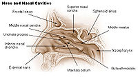 There are four sinuses dealt with by means of this surgery: The frontal sinuses located in the forehead, the maxillary sinuses in the cheeks, the ethmoid sinuses between the eyes, and finally the sphenoid sinuses located in the back of the nasal cavity at the base of the skull.
Controversy exists as to whether or not the maxillary ostium should be enlarged or not depending on the disease status of the maxillary sinus. However, the medical literature would support a wide antrostomy and complete clearance down to healthy mucosa if fungal mucin is present within the sinus. In this circumstance, the ostium is enlarged superiorly to orbital floor and posteriorly to posterior fontanelle to allow wide access for clearance.
 Complete maxillary debridement can be accomplished via either trans-ostial clearance which can be quite tedious. A newer technique, canine fossa trephination, can accomplish this same task faster and with few side effects.
Endoscopic access to pituitary tumors has been found to be quite useful as well. Using endoscopes for hypophysectomy allows excellent visualization within the sella and more complete tumor removal than would be available via microsurgical technique.
This can be divided into:
You may classify to
This tells the surgeon what neighboring structures are located next to the probe to guide dissection without invading unintended structures. Bleeding and anatomic variants can also alter a surgeon's view of landmarks through an endoscope. Hence, CT-navigation assistance in sinus surgery is used to improve anatomical identification and avoid damage to vital structures such as the brain and eyes.
Definitive proof that CT-Navigation improves outcomes and decreases complications is still lacking. A Swedish study of 212 patients undergoing sphenoethmoidectomy published in 2008 concluded the additional benefit from the use of CT-Navigation.
Fovea Ethmoidalis is the true roof of the ethmoid cavities and the bony marker of the skull base. It extends from the orbital lamellae laterally to the lateral lamella of the cribriform medially.
Sinusitis
Sinusitis is inflammation of the paranasal sinuses, which may be due to infection, allergy, or autoimmune issues. Most cases are due to a viral infection and resolve over the course of 10 days...
and nasal polyps, including bacterial, fungal, recurrent acute, and chronic sinus problems. Ample research supports its record of safety and success.
FESS is a relatively recent surgical procedure that uses nasal endoscopes (using Hopkins rod lens technology) through the nostrils to avoid cutting the skin. These endoscopes have diameters of 4mm (adult use) and 2.7mm (pediatric use) and come in varying angles of view from 0 degrees to 30, 45, 70, 90, and 120 degrees. They provide good illumination of the inside of the nasal cavity and sinuses.
FESS came into existence because of pioneering work of Messerklinger and Stamberger (Graz, Austria.) Other surgeons have made additional contributions (first published in USA by Kennedy in 1985).
Technique
Maxillary sinus
One of the most accepted means of functionally enlarging the maxillary ostium is to perform an uncinectomy via the "swing door" technique. This initially removes the vertical process of the uncinate via backbiter inferiorly and sickle knife superiorly. The uncinate is swung medially and then severed at its lateral attachment. This is followed by a submucosal removal of the horizontal process of the uncinate and subsequent trimming of the mucosa to fully visualize the maxillary os.Controversy exists as to whether or not the maxillary ostium should be enlarged or not depending on the disease status of the maxillary sinus. However, the medical literature would support a wide antrostomy and complete clearance down to healthy mucosa if fungal mucin is present within the sinus. In this circumstance, the ostium is enlarged superiorly to orbital floor and posteriorly to posterior fontanelle to allow wide access for clearance.
Extended approaches
More recently, the paranasal sinuses have been found to be a relatively low-morbidity approach to selected tumors of the anterior and posterior cranial fossa.Endoscopic access to pituitary tumors has been found to be quite useful as well. Using endoscopes for hypophysectomy allows excellent visualization within the sella and more complete tumor removal than would be available via microsurgical technique.
This can be divided into:
- approaches to the anterior cranial fossa
- approaches to the mid cranial fossa
- approaches to the posterior cranial fossa
- access to the infratemporal fossa (incl. pterygopalatine fissure)
- access to the sella turcica
- orbital access
- optic nerve access
Complications
Extreme care is required with this surgery due to the paranasal sinus' proximity to the orbits, brain, internal carotid arteries, and optic nerves. However, even with these possible serious risks, there are many benefits to be reaped by a patient with appropriate indications from a well-performed ESS. As the degree of difficulty increases with these surgeries, a surgeon with appropriate experience must be present to manage the procedure. This is especially true in approaches to neurosurgical procedures.You may classify to
- Orbital complication: including orbital haemorrhage, abscess, damage to optic nerve
- Intracranial complication: including CSF Leak, meningitis, Brain abscess, Intracranial haemorrhage
- Nasal complication: including adhesion formation, anosmia, hyposmia, injury to lacrimal duct
The use of CT-Navigation in Endoscopic Sinus Surgery
A Computed Tomography (CT)Navigation system is a tool that is used by surgeons to better correlate surgical anatomy with pre-operative CT imaging. A computer is used to identify the 3-dimensional location of a probe tip placed within the patient's nose or sinuses. The computer will then identify the spot on the CT image where the surgeons probe is placed.This tells the surgeon what neighboring structures are located next to the probe to guide dissection without invading unintended structures. Bleeding and anatomic variants can also alter a surgeon's view of landmarks through an endoscope. Hence, CT-navigation assistance in sinus surgery is used to improve anatomical identification and avoid damage to vital structures such as the brain and eyes.
Definitive proof that CT-Navigation improves outcomes and decreases complications is still lacking. A Swedish study of 212 patients undergoing sphenoethmoidectomy published in 2008 concluded the additional benefit from the use of CT-Navigation.
Anatomy
Planum Sphenoidale marks the posterior limit of the anterior skull base. This bony structure is the plane created by the medial confluence of the lesser wing of the sphenoid bone.Fovea Ethmoidalis is the true roof of the ethmoid cavities and the bony marker of the skull base. It extends from the orbital lamellae laterally to the lateral lamella of the cribriform medially.