

Eosinophilic
Encyclopedia
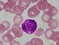
Eosinophilic refers to the stain
Stain
A stain is a discoloration that can be clearly distinguished from the surface, material, or medium it is found upon. Stains are caused by the chemical or physical interaction of two dissimilar materials...
ing of certain tissues
Biological tissue
Tissue is a cellular organizational level intermediate between cells and a complete organism. A tissue is an ensemble of cells, not necessarily identical, but from the same origin, that together carry out a specific function. These are called tissues because of their identical functioning...
, cells
Cell (biology)
The cell is the basic structural and functional unit of all known living organisms. It is the smallest unit of life that is classified as a living thing, and is often called the building block of life. The Alberts text discusses how the "cellular building blocks" move to shape developing embryos....
, or organelle
Organelle
In cell biology, an organelle is a specialized subunit within a cell that has a specific function, and is usually separately enclosed within its own lipid bilayer....
s after they have been washed with eosin
Eosin
Eosin is a fluorescent red dye resulting from the action of bromine on fluorescein. It can be used to stain cytoplasm, collagen and muscle fibers for examination under the microscope. Structures that stain readily with eosin are termed eosinophilic....
, a dye
Dye
A dye is a colored substance that has an affinity to the substrate to which it is being applied. The dye is generally applied in an aqueous solution, and requires a mordant to improve the fastness of the dye on the fiber....
.
Eosin is an acidic dye; thus, the structure being stained is basic
Base (chemistry)
For the term in genetics, see base A base in chemistry is a substance that can accept hydrogen ions or more generally, donate electron pairs. A soluble base is referred to as an alkali if it contains and releases hydroxide ions quantitatively...
.
Eosinophilic describes the appearance of cells and structures seen in histological section
Histological section
Histological section refers to thin slices of tissue applied to a microscopic slide, usually around 5 to 10 micrometres thick, which are viewed under a microscope...
s that take up the staining dye eosin
Eosin
Eosin is a fluorescent red dye resulting from the action of bromine on fluorescein. It can be used to stain cytoplasm, collagen and muscle fibers for examination under the microscope. Structures that stain readily with eosin are termed eosinophilic....
. This is a bright-pink dye that stains the cytoplasm of cells, as well as extracellular proteins such as collagen
Collagen
Collagen is a group of naturally occurring proteins found in animals, especially in the flesh and connective tissues of mammals. It is the main component of connective tissue, and is the most abundant protein in mammals, making up about 25% to 35% of the whole-body protein content...
.
Such eosinophilic structures are, in general, composed of protein
Protein
Proteins are biochemical compounds consisting of one or more polypeptides typically folded into a globular or fibrous form, facilitating a biological function. A polypeptide is a single linear polymer chain of amino acids bonded together by peptide bonds between the carboxyl and amino groups of...
.
The stain eosin is usually combined with a stain called hematoxylin to produce a hematoxylin and eosin-stained section
Histological section
Histological section refers to thin slices of tissue applied to a microscopic slide, usually around 5 to 10 micrometres thick, which are viewed under a microscope...
(also called an H&E stain
H&E stain
H&E stain, HE stain or hematoxylin and eosin stain is a popular staining method in histology. It is the most widely used stain in medical diagnosis; for example when a pathologist looks at a biopsy of a suspected cancer, the histological section is likely to be stained with H&E and termed H&E...
, HE or H+E section). This is the most widely-used histological stain in medical diagnosis; for example, when a pathologist examines a biopsy
Biopsy
A biopsy is a medical test involving sampling of cells or tissues for examination. It is the medical removal of tissue from a living subject to determine the presence or extent of a disease. The tissue is generally examined under a microscope by a pathologist, and can also be analyzed chemically...
of a suspected cancer, the biopsy will have been stained with H&E.
Some structures seen inside cells are described as being eosinophilic, for example, Lewy bodies
Lewy body
Lewy bodies are abnormal aggregates of protein that develop inside nerve cells in Parkinson's disease , Lewy Body Dementia and some other disorders. They are identified under the microscope when histology is performed on the brain....
, Mallory bodies
Mallory body
In histopathology, a Mallory body, Mallory-Denk body, and Mallory's hyaline, is an inclusion found in the cytoplasm of liver cells.-Associated conditions:...
.
See also
- basophilicBasophilicBasophilic is a technical term used by histologists. It describes the microscopic appearance of cells and tissues, as seen down the microscope, after a histological section has been stained with a basic dye. The most common such dye is haematoxylin....
(affinity to hematoxyllnHaematoxylinHaematoxylin, hematoxylin, Natural Black 1, or C.I. 75290 is extracted from the heartwood of the logwood tree. When oxidized it forms haematein, a compound that forms strongly coloured complexes with certain metal ions, the most notable ones being Fe and Al salts. Metal-haematein complexes are used...
) - EosinophiliaEosinophiliaEosinophilia is a condition in which the eosinophil count in the peripheral blood exceeds 0.45×10⁹/L . A marked increase in non-blood tissue eosinophil count noticed upon histopathologic examination is diagnostic for tissue eosinophilia. Several causes are known, with the most common being...
- Eosinophilic Meningitis