
Calcaneal fracture
Encyclopedia
Calcaneal fracture, also known as Lover's fracture and Don Juan fracture, is a fracture of the calcaneus. It is usually caused by a fall from height when one lands on his or her feet. These fractures represent approximately 2% of all fractures but 60% of tarsal bone fractures. The name lover's fracture is derived from the fact that a lover may jump from great heights while trying to escape from the lover's spouse.
The calcaneus, also known as the heel bone, is the largest of the tarsal bones and articulates with the cuboid bone anteriorly and the talus bone superiorly. It is responsible for transmitting the majority of the body's weight from the talus bone to the ground.
Calcaneal fractures are categorized into two types: Intra- and Extrarticular fractures on the basis of subtalar joint involvement. Intrarticular fractures are more common and involve the posterior talar articular facet of the calcaneus. The Sanders system classifies these fractures into four types, based on the location of the fracture at the posterior articular surface. Extrarticular fractures are less common, and located anywhere outside the the subtalar joint. Extrarticular fractures are categorized depending on whether the involvement of the calcaneus is anterior (Type A), Middle (Type B) or Posterior (Type C).
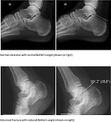
The Angle of Gissane, or "Critical Angle", is the angle formed by the downward and upward slopes of the calcaneal superior surface. On a lateral radiograph, an angle of Gissane of > 130° suggests fracture of the posterior subtalar joint surface.[3] The Böhler's angle, or "Tuber Angle", is another normal anatomic landmark seen in lateral radiographs. It is formed by the intersection of 1) a line from the highest point of the posterior articular facet to the highest point of the posterior tuberosity, and 2) a line from the former to the highest point on the anterior articular facet. An angle < 20° suggests a depression of posterior facet and possible calcaneal fracture.
The Sanders Classification system is the most commonly used system for categorizing intrarticular fractures. There are 4 types:
- Type I fractures are non-displaced fractures (displacement < 2 mm).
- Type II fractures consist of a single intrarticular fracture that divides the calcaneus into 2 pieces.
--Type IIA → fracture occurs on lateral aspect of calcaneus.
--Type IIB → fracture occurs on central aspect of calcaneus.
--Type IIC → fracture occurs on medial aspect of calcaneus.
- Type III fractures consist of two intrarticular fractures that divide the calcaneus into 3 articular pieces.
--Type IIIAB → two fracture lines are present, one lateral and one central.
--Type IIIAC → two fracture lines are present, one lateral and one medial.
--Type IIIBC → two fracture lines are present, one central and one medial.
- Type IV fractures consist of fractures with more than three intrarticular fractures.
Extrarticular fractures include all fractures that do not involve the posterior facet of the subtalar joint.
- Type A → involve the anterior calcaneus
- Type B → involve the middle calcaneus. This includes the sustentaculum tali, trochlear process and lateral process.
- Type C → involve the posterior calcaneus, the posterior tuberosity and medial tubercle included.
Displaced intrarticular fractures require surgical intervention within 3 weeks of fracture, before bone consolidation has ocurred. Conservative surgery consists of closed reduction with percutaneous fixation. This technique is associated with less wound complications, better soft tissue healing (because of less soft tissue manipulation) and decreased intraoperative time. However, this procedure has increased risk of inadequate calcaneal bone fixation, compared to open procedures. Currently, open reduction with internal fixation (ORIF) is usually the preferred surgical approach when dealing with displaced intrarticular fractures. Newer, more innovative surgical techniques and equipment have decreased the incidence of intra- and post-operative complications.
The calcaneus, also known as the heel bone, is the largest of the tarsal bones and articulates with the cuboid bone anteriorly and the talus bone superiorly. It is responsible for transmitting the majority of the body's weight from the talus bone to the ground.
Calcaneal fractures are categorized into two types: Intra- and Extrarticular fractures on the basis of subtalar joint involvement. Intrarticular fractures are more common and involve the posterior talar articular facet of the calcaneus. The Sanders system classifies these fractures into four types, based on the location of the fracture at the posterior articular surface. Extrarticular fractures are less common, and located anywhere outside the the subtalar joint. Extrarticular fractures are categorized depending on whether the involvement of the calcaneus is anterior (Type A), Middle (Type B) or Posterior (Type C).
The Angle of Gissane, or "Critical Angle", is the angle formed by the downward and upward slopes of the calcaneal superior surface. On a lateral radiograph, an angle of Gissane of > 130° suggests fracture of the posterior subtalar joint surface.[3] The Böhler's angle, or "Tuber Angle", is another normal anatomic landmark seen in lateral radiographs. It is formed by the intersection of 1) a line from the highest point of the posterior articular facet to the highest point of the posterior tuberosity, and 2) a line from the former to the highest point on the anterior articular facet. An angle < 20° suggests a depression of posterior facet and possible calcaneal fracture.
Clinical Presentation
The most common symptom is pain over the heel area, especially when the heel is palpated or squeezed. Patients usually have a history of recent trauma to the area or fall from a height. Other symptoms include: inability to bear weight over the involved foot, limited mobility of the foot, and limping. Upon inspection, the examiner may notice swelling, redness, and hematomas. A hematoma extending to the sole of the foot is called "Mondor Sign", and is pathognomonic for calcaneal fracture. The heel may also become widened with associated edema due to displacement of lateral calcaneal border. Involvement of soft tissue (tendons, skin, etc,) should be evaluated because soft tissue injury has been associated to serious complications (see below).Diagnosis
Conventional radiography is usually the initial assessment tool when calcaneal fracture is suspected. Recommended x-ray views are (a) axial, (b) anteroposterior, (C) oblique views and (d) views with dorsiflexion and internal rotation of the foot. However, conventional radiography is limited for visualization of calcaneal anatomy, especially at the subtalar joint. CT Scan is currently the imaging study of choice for evaluating calcaneal injury and has substituted conventional radiography in the classification of calcaneal fractures. Axial and coronal views are taken for proper visualization of the calcaneus, subtalar, calcaneocuboid and talonavicular joints.The Sanders Classification system is the most commonly used system for categorizing intrarticular fractures. There are 4 types:
- Type I fractures are non-displaced fractures (displacement < 2 mm).
- Type II fractures consist of a single intrarticular fracture that divides the calcaneus into 2 pieces.
--Type IIA → fracture occurs on lateral aspect of calcaneus.
--Type IIB → fracture occurs on central aspect of calcaneus.
--Type IIC → fracture occurs on medial aspect of calcaneus.
- Type III fractures consist of two intrarticular fractures that divide the calcaneus into 3 articular pieces.
--Type IIIAB → two fracture lines are present, one lateral and one central.
--Type IIIAC → two fracture lines are present, one lateral and one medial.
--Type IIIBC → two fracture lines are present, one central and one medial.
- Type IV fractures consist of fractures with more than three intrarticular fractures.
Extrarticular fractures include all fractures that do not involve the posterior facet of the subtalar joint.
- Type A → involve the anterior calcaneus
- Type B → involve the middle calcaneus. This includes the sustentaculum tali, trochlear process and lateral process.
- Type C → involve the posterior calcaneus, the posterior tuberosity and medial tubercle included.
Treatment
Non-surgical treatment is indicated for extrarticular fractures and Sanders Type I intrarticular fractures, provided that the calcaneal weight-bearing surface and foot function are not compromised. Physicians may choose to perform closed reduction with or without fixation (casting), or fixation alone (without reduction), depending on the individual case. Recommendations include no weight-bearing for a few weeks followed by range-of-motion exercises and progressive weight bearing for a period of 2-3 months.Displaced intrarticular fractures require surgical intervention within 3 weeks of fracture, before bone consolidation has ocurred. Conservative surgery consists of closed reduction with percutaneous fixation. This technique is associated with less wound complications, better soft tissue healing (because of less soft tissue manipulation) and decreased intraoperative time. However, this procedure has increased risk of inadequate calcaneal bone fixation, compared to open procedures. Currently, open reduction with internal fixation (ORIF) is usually the preferred surgical approach when dealing with displaced intrarticular fractures. Newer, more innovative surgical techniques and equipment have decreased the incidence of intra- and post-operative complications.