
Articular capsule of the knee joint
Encyclopedia
The articular capsule of the knee joint (commonly referred to as capsular ligament
) is wide and lax; thin in front and at the side; and contains the patella ("knee cap"), ligament
s, menisci
, and bursae. The capsule consists of a synovial and a fibrous membrane
separated by fatty deposits anteriorly and posteriorly.
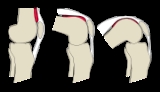
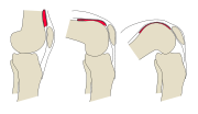 Anteriorly, the reflection of the synovial membrane lies on the femur
; located at some distance from the cartilage because of the presence of the suprapatellar bursa. Above, the reflection appears lifted from the bone by underlying periosteal
connective tissue
. In a standing posture, the suprapatellar bursa is seemingly redundant. It is however also referred to as the suprapatellar synovial recess as it gradually unfolds as the knee is flexed; to open up completely when the knee is flexed 130 degrees. The suprapatellar bursa is prevented from being pinched during extension by the articularis genu muscle
. On the tibia
, the anterior reflection and attachment of the synovial membrane is located near the cartilage.
Anteriorly, the infrapatellar fat pad is inserted below the patella and between the two membranes. It extends from the lower margin of the patella above, to the infrapatellar synovial fold below. With its free upper margin, this fold extends dorsally through the joint space to surround the two cruciate ligaments from the front, thus dividing the surrounding joint space into two chambers. Laterally of this are a pair of alar folds.
Posteriorly, the femoral attachment of the synovial membrane is located at the cartilaginous margin of the lateral
and medial femoral condyle
s, why the joint space has two dorsal extensions. Between these, the synovial membrane passes in front of the anterior
and posterior cruciate ligament
s, why these ligaments are both intracapsular and extra-articular with their tibial attachment located exactly on the cartilage margin. Both the lateral
and medial meniscus
are, however, located within the synovial capsule.
Above and in front, beneath the tendon of the Quadriceps femoris, it is represented only by the synovial membrane.
Its chief strengthening bands are derived from the fascia lata
and from the tendon
s surrounding the joint.
Adding to the complex structure of the knee space, there are remnants of three embryonic septal
divisions of the knee space called synovial plicae:
Ligament
In anatomy, the term ligament is used to denote any of three types of structures. Most commonly, it refers to fibrous tissue that connects bones to other bones and is also known as articular ligament, articular larua, fibrous ligament, or true ligament.Ligament can also refer to:* Peritoneal...
) is wide and lax; thin in front and at the side; and contains the patella ("knee cap"), ligament
Ligament
In anatomy, the term ligament is used to denote any of three types of structures. Most commonly, it refers to fibrous tissue that connects bones to other bones and is also known as articular ligament, articular larua, fibrous ligament, or true ligament.Ligament can also refer to:* Peritoneal...
s, menisci
Meniscus (anatomy)
In anatomy, a meniscus is a crescent-shaped fibrocartilaginous structure that, in contrast to articular disks, only partly divides a joint cavity. In humans it is present in the knee, acromioclavicular, sternoclavicular, and temporomandibular joints; in other organisms they may be present in other...
, and bursae. The capsule consists of a synovial and a fibrous membrane
Collagen
Collagen is a group of naturally occurring proteins found in animals, especially in the flesh and connective tissues of mammals. It is the main component of connective tissue, and is the most abundant protein in mammals, making up about 25% to 35% of the whole-body protein content...
separated by fatty deposits anteriorly and posteriorly.
Synovial membrane
Femur
The femur , or thigh bone, is the most proximal bone of the leg in tetrapod vertebrates capable of walking or jumping, such as most land mammals, birds, many reptiles such as lizards, and amphibians such as frogs. In vertebrates with four legs such as dogs and horses, the femur is found only in...
; located at some distance from the cartilage because of the presence of the suprapatellar bursa. Above, the reflection appears lifted from the bone by underlying periosteal
Periosteum
Periosteum is a membrane that lines the outer surface of all bones, except at the joints of long bones. Endosteum lines the inner surface of all bones....
connective tissue
Connective tissue
"Connective tissue" is a fibrous tissue. It is one of the four traditional classes of tissues . Connective Tissue is found throughout the body.In fact the whole framework of the skeleton and the different specialized connective tissues from the crown of the head to the toes determine the form of...
. In a standing posture, the suprapatellar bursa is seemingly redundant. It is however also referred to as the suprapatellar synovial recess as it gradually unfolds as the knee is flexed; to open up completely when the knee is flexed 130 degrees. The suprapatellar bursa is prevented from being pinched during extension by the articularis genu muscle
Articularis genu muscle
The articularis genu is a small skeletal muscle located anteriorly on the thigh just above the knee.- Origin and insertion :...
. On the tibia
Tibia
The tibia , shinbone, or shankbone is the larger and stronger of the two bones in the leg below the knee in vertebrates , and connects the knee with the ankle bones....
, the anterior reflection and attachment of the synovial membrane is located near the cartilage.
Anteriorly, the infrapatellar fat pad is inserted below the patella and between the two membranes. It extends from the lower margin of the patella above, to the infrapatellar synovial fold below. With its free upper margin, this fold extends dorsally through the joint space to surround the two cruciate ligaments from the front, thus dividing the surrounding joint space into two chambers. Laterally of this are a pair of alar folds.
Posteriorly, the femoral attachment of the synovial membrane is located at the cartilaginous margin of the lateral
Lateral condyle of femur
The lateral condyle is one of the two projections on the lower extremity of femur. It is the more prominent and is the broader both in its antero-posterior and transverse diameters....
and medial femoral condyle
Medial condyle of femur
The medial condyle is one of the two projections on the lower extremity of femur.The medial condyle is larger than the lateral condyle due to more weight bearing caused by the center of gravity being medial to the knee. On the posterior surface of the condyle the linea aspera turns into the...
s, why the joint space has two dorsal extensions. Between these, the synovial membrane passes in front of the anterior
Anterior cruciate ligament
The anterior cruciate ligament is a cruciate ligament which is one of the four major ligaments of the human knee. In the quadruped stifle , based on its anatomical position, it is referred to as the cranial cruciate ligament.The ACL originates from deep within the notch of the distal femur...
and posterior cruciate ligament
Posterior cruciate ligament
The posterior cruciate ligament is one of the four major ligaments of the knee. It connects the posterior intercondylar area of the tibia to the medial condyle of the femur...
s, why these ligaments are both intracapsular and extra-articular with their tibial attachment located exactly on the cartilage margin. Both the lateral
Lateral meniscus
The lateral meniscus, also called the external semilunar fibrocartilage, is a fibrocartilaginous band that spans the lateral side of the interior of the knee joint. It is one of two menisci of the knee, the other being the medial meniscus. It is nearly circular and covers a larger portion of the...
and medial meniscus
Medial meniscus
-External links: *...
are, however, located within the synovial capsule.
Fibrous membrane
It is a thin, but strong, fibrous membrane which is strengthened in almost its entire extent by bands inseparably connected with it.Above and in front, beneath the tendon of the Quadriceps femoris, it is represented only by the synovial membrane.
Its chief strengthening bands are derived from the fascia lata
Fascia lata
-Thickness:It is an investment for the whole of the thigh, but varies in thickness in different parts.Thus, it is thicker in the upper and lateral part of the thigh, where it receives a fibrous expansion from the Glutæus maximus, and where the Tensor fasciæ latæ is inserted between its layers; it...
and from the tendon
Tendon
A tendon is a tough band of fibrous connective tissue that usually connects muscle to bone and is capable of withstanding tension. Tendons are similar to ligaments and fasciae as they are all made of collagen except that ligaments join one bone to another bone, and fasciae connect muscles to other...
s surrounding the joint.
Bursae
The numerous bursae surrounding the knee joint can be divided into the communicating and the non-cummunicating bursae:- Communicating bursae:
- The suprapatellar bursa, the largest bursa, extends the joint space anteriorly and proximally.
- The subpopliteal recess and semimembranosus bursa are located posteriorly and are much smaller
- The lateral and medial subtendinous bursae of gastrocnemius are located at the origin of the two heads of the gastrocnemius muscleGastrocnemius muscleIn humans, the gastrocnemius muscle is a very powerful superficial pennate muscle that is in the back part of the lower leg. It runs from its two heads just above the knee to the heel, and is involved in standing, walking, running and jumping. Along with the soleus muscle it forms the calf muscle...
.
- Non-communicating bursae:
- The subcutaneous prepatellar bursa is located in front of the patella.
- The [deep] infrapatellar bursa is located under the patella, between the patellar ligament and the fibrous membrane of the joint capsule. It is communicating with the joint space in particular cases.
- Other less regularly present bursae include the subfascial prepatellar, the subtendinous prepatellar, and the subcutaneous prepatellar bursae.
Adding to the complex structure of the knee space, there are remnants of three embryonic septal
Septum
In anatomy, a septum is a wall, dividing a cavity or structure into smaller ones.-In human anatomy:...
divisions of the knee space called synovial plicae:
- The suprapatellar plica dividing the suprapatellar recess
- The infrapatellar plica, in front of the anterior cruciate ligamentAnterior cruciate ligamentThe anterior cruciate ligament is a cruciate ligament which is one of the four major ligaments of the human knee. In the quadruped stifle , based on its anatomical position, it is referred to as the cranial cruciate ligament.The ACL originates from deep within the notch of the distal femur...
, reaches from the intercondylar notch to the infrapateller fat pad - The medial patellar plica, located adjacent to the patella's medial facet, runs vertically along the medial joint capsule